What's happened
Skin cancer is a malignant tumor. The disease is rare and is detected in only 0.3 percent of cases.
Sometimes there is a transmission of oncopathology by inheritance from mother to child. In this case, the formation of a malignant process of congenital type is noted. In other words, the neoplasm is integrated into the child’s body, facilitated by genetic congruence.
There are often situations when the disease begins to behave in an unpredictable way, that is, cases have been recorded in which a complete cure occurred.
In addition, the disease is characterized by a change in the growth rate of tumor formation. Sometimes the pathological process is accompanied by rapid development, and it happens that phenomena that are aggressive in nature become passive.
Types of tumors on the arm under the skin
- Benign tumors of the upper extremities
- Malignant tumors of the hand
A tumor on the arm under the skin can be benign or malignant.
New growths develop on the finger, hand, shoulder, forearm, and wrist. The main sign of a skin tumor is that at an early stage, upon palpation, it becomes clear that it is not associated with bone formations.
If the structure of the neoplasm is homogeneous, it can be assumed to be benign.
If the density is uneven and there are areas of softening, this indicates tissue necrosis inside the tumor and may be a sign of cancer.
The simplest classification of neoplasms on the upper extremities:
- benign – can be located on soft tissues and bones;
- malignant without metastasis - the location is similar;
- metastatic tumors - the same location.
Benign tumors of the upper extremities
Bone:
- Cysts. This neoplasm affects the finger and is localized in the phalanx area. Causes pain, joint mobility is limited.
- Chondromas and endrochondromas. They are localized in the area of the phalanges, formed from cartilaginous cells, can be located inside or outside the bone, be single or multiple. If the tumors are multiple, there is a risk of malignant degeneration.
Benign soft tissue tumors.
- Cysts. Epidermoid and implantation. They occur when the surface epithelium penetrates into the deep layers of the skin. Causes of appearance: trauma, amputation.
- Xanthomas. They occur near the fingers on the side of the palm and cause pathological division of epithelial cells of tendon sheaths, sweat and sebaceous glands, blood vessels and lymphatic ducts.
- Fibroids are pronounced skin changes. They are located on the inside of the fingers and palm and can cause acute pain when pressed.
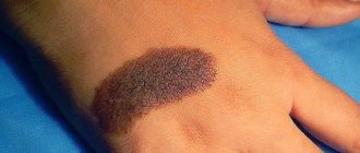
- Pigmented. They rise above the skin and may have hair. There is a possibility of degeneration into melosarcoma.
- Tendon ganglia are one of the more private formations on the hand that appear after injury or with constant physical activity. They are localized above the projection of the tendons and have an elongated shape.
- Warts. Occur when the epithelium is infected with the papilloma virus. Can be solitary or growing. There is a risk of malignant degeneration.
- Lipomas are tumors of fatty tissue. They can grow through the bones and turn the hand into something like a pillow. Localized at the initial stage on the palm.
- Hygromas are formed from the joint capsule. In the initial stage, when pressure is applied, the neoplasm goes into the joint cavity.
- Glomus tumor is a neoplasm of the circulatory system. Localized in the wrist and forearm, it is extremely painful. Nerve segments and arterial anastomosis are involved in its formation. There is a risk of progression to cancer.
- Hemangiomas - vascular tumors - are congenital. In 80% of cases it has a doughy consistency, but sometimes it is hard. Most often located at the base of the thumb. There is severe pain when pressed.
- Sarcomas. They are rare, at the initial stage of development they resemble fibroids, and are localized in the connective tissue - fascia. They quickly increase, causing acute pain, deforming and destroying the bone.
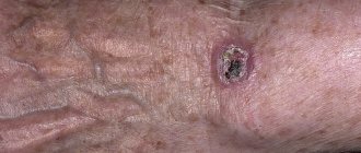
- Skin cancer may initially be confused with a dermatological disease. Neoplasms that rise above the skin surface - most often on the hand - ulcerate, forming erosions with raised edges. Purulent neoplasms become infected and an unpleasant odor appears. Subsequently they grow deeper into the tissues.
- Synomvioma is a soft neoplasm with unclear contours that appears with pathological changes in the articular sheaths and capsules. Metastases are rare, with an increased tendency to relapse. May be benign.
- Angiosarcoma. The symptom is the appearance of dark spots on the skin, then turning into nodular vascular formations.
- Metastatic tumors are more common on the fingers. They have no external signs.
New growths on the hand are difficult to miss.
When diagnosed at the initial stage, the transformation of a tumor into a malignant one or the development of cancer can be avoided.
Any tumor on the hand, even if it does not cause pain, is a reason to consult a doctor for diagnosis. The sooner treatment begins, the lower the likelihood of degeneration or metastasis.
Source: //upraznenia.ru/opuxol-na-ruke-pod-kozhej.html
Classification
Skin cancer in childhood comes in several varieties.
Melanoma
This form of cancer is considered one of the most dangerous. It is characterized by rapid growth and multiple spread of metastases to lymph nodes and other anatomical structures.
Melanoma is rarely diagnosed in children and accounts for about 0.3 percent of all malignant tumors. However, as practice shows, cases of cancer diagnosis are increasing every year.
Pigment cells of the skin take part in the formation of melanoma. The peak of the disease occurs at ages 4 to 6 years and 11 to 15 years. In most cases, the upper and lower extremities, neck, head and torso are affected.
In addition, melanoma is classified into local (no secondary lesions) and widespread.
Basal cell
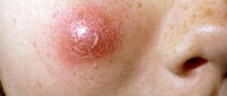
This type of cancer is also called basal cell carcinoma. The location of the neoplasm is the child’s face. The tumor has a dense consistency. Outwardly it resembles a nodular formation. A characteristic feature is its fusion with the skin or a slight elevation above its surface.
As the oncological process progresses, the number of nodes may increase. In addition, they are able to merge into one large spot.
The growth of a malignant tumor occurs at a slow pace. Often the pathology can occur for a long time without the manifestation of any clinical signs.
On this topic
- Oncodermatology
Prognosis for skin basal cell carcinoma
- Natalya Gennadievna Butsyk
- December 10, 2020
As it grows, other tissues and anatomical structures close to the affected area begin to become involved in the process. Bone, cartilage, muscle tissue and mucous tissues may also be affected. If the tumor grows into the mucous layers, there is a sharp deterioration in the child’s well-being.
Distant metastasis is rare. In most cases, the lymph nodes are affected.
Squamous
The site of localization is often the face, ears, scalp, legs and arms. Also, the external genitalia may be involved in the oncological process. In children, the cause of this type of tumor in most cases is the development of xeroderma pigmentosum. This is a rare pathology characterized by skin rashes.
Squamous cell carcinoma is characterized by rapid development compared to the type described above. Also, the neoplasm can metastasize to nearby lymph nodes, but this is rare. In approximately 35 percent of cases, parents and children turn to specialists when the disease has already reached the last stage of development.
Signs of malignant lumps
The formations that form under the skin on the palms can have different origins. A malignant tumor can be recognized by a number of signs:
- The lump does not have clear and even edges. But at the first stage it is imperceptible, in addition, there is no discomfort, pain or other symptoms.
- Tumor growth, which is often accompanied by fever and general malaise. If the lump has grown more than a centimeter and is causing discomfort, urgent medical attention is needed.
- Mobility is not typical for a malignant tumor. As a rule, they grow into the skin, so pain is felt upon palpation. In severe cases, blood or pus may appear from the lump.
- Malignant tumors often cause fever. For a long time the temperature can remain at thirty-seven degrees, but then rises, reaching forty. This is caused by inflammation of the lymph nodes.
Stages
Like most cancers, skin cancer in a child goes through several stages of formation.
Zero
Abnormal cells are just beginning to form on the surface of the skin. In this case, it is difficult to suspect the disease. As a rule, it is discovered accidentally during diagnosis due to another pathology. The prognosis is favorable. Complete cure can be achieved in 100% of cases.
First
The size of the tumor reaches no more than two centimeters. There is a gradual germination into the deeper layers of the skin.
There are still no metastases. If you choose the right therapeutic tactics, the disease can be cured.
Second
The diameter of the neoplasm can be from 2.5 to 5 centimeters. Growing into all layers of the dermis is noted. The pathological process is accompanied by burning, itching and pain. A single secondary lesion begins to form in the lymph node.
Third
The tumor is growing and is already more than five centimeters in diameter. In this condition, the patient experiences discomfort. The surface of the neoplasm becomes covered with ulcerations. Metastases spread to cartilage and bone tissue.
The body temperature begins to rise and the patient’s general well-being worsens. In addition, metastasis to regional lymph nodes is observed.
Fourth
The compaction reaches impressive sizes. It has uneven outlines, and crusts and ulcers form on the surface.
This condition is accompanied by severe intoxication. Metastases affect vital organs and are found in the liver, lungs and bones.
Description
Blastoma is a pathological proliferation of tissues, the structure of which consists of deformed cells. These are elements that have lost their normal functioning and form. Their distinctive feature is that even if they are no longer negatively affected, they still continue to multiply and contribute to the formation of blastoma.
On this topic
- General
What is a cancer examination
- Natalya Gennadievna Butsyk
- December 6, 2020
Tumors can be of two types - benign and malignant. They differ significantly from each other. First of all, we are talking about the fact that the first type is characterized by the growth of neoplasms within the affected organ, and the adjacent tissues only move apart.
If we talk about oncological processes, then in this case the tumor will grow deep into the tissues, resulting in damage and destruction of blood vessels, which leads to the spread of cancer cells throughout the body through the bloodstream. This is how metastasis begins, which is how benign blastomas differ from malignant ones.
Blastoma in its formation goes through several stages of development:
- first - pathological cells begin to repeatedly divide and increase;
- second – the lesion is growing;
- third – the formation of a benign neoplasm occurs;
- fourth – the process of malignancy of the tumor is noted.
Subsequently, the tumor grows and does not respond to the regulatory systems of the human body. If therapeutic measures are not taken in a timely manner, metastases will form, which leads to even more serious problems and complicates treatment.
Causes
To date, the exact triggering factors contributing to the development of skin cancer in children have not been established. However, according to most experts, genetic pathologies such as Fanconi anemia, Down syndrome and others are given first place.
In addition, among the possible causes of the appearance of a malignant tumor may be prolonged exposure to ultraviolet rays or ionizing radiation. Also no less important is the decrease in the child’s immune system and the presence of age spots, which begin to progress with constant trauma and degenerate into oncological tumors.
How to avoid getting skin cancer? What to avoid?
Sunlight. The most proven cause of both types of skin cancer, as well as melanoma, is exposure to sunlight. If you like to travel to hot countries, have fair hair and skin, or your work involves prolonged exposure to the sun, you should seriously consider UV protection.
Precancerous skin diseases are the next factor that may precede the development of the squamous cell form: actinic (solar) keratoses and cheilitis, leukoplakia, human papillomavirus infection of the mucous membranes and genitals. This type of tumor can also develop against the background of scar changes after burns or radiation therapy.
Contact with carcinogens
Various chemicals can lead to the development of skin cancer: arsenic and petroleum products.
Weakened immune system. People taking immunosuppressive drugs after an organ transplant or people living with HIV have an increased risk of developing squamous cell skin cancer.
Symptoms
The development of cancer will primarily be indicated by a change in the shade of the pigment formation. In addition, the tumor grows rapidly and rises slightly above the surface of the skin. There is rapid growth of the affected area.
The lump may bleed and ulcerate. Cracks form in the affected area. If a pigmented nevus has hairs, they fall out over time.
Also, the beginning of the development of the pathological process is characterized by inflammation of the corolla, which surrounds the pathological area.
On this topic
- Oncodermatology
Prognosis for skin cancer at various stages
- Natalya Gennadievna Butsyk
- December 6, 2020
The formation of a tumor may be accompanied by burning, pain and itching.
As the cancer progresses, the clinical picture is complemented by the spread of metastases to nearby anatomical structures, and the lymph nodes become enlarged.
Next, metastases begin to affect distant organs and tissues, which causes general intoxication of the patient’s body.
External signs of damage to the digestive system
Red spots on the body with stomach cancer occur due to a decrease in the immune properties of the body. Nodular thickenings are observed in various parts of the body. Dangerous cells spread throughout the body, forming brownish areas. Black formations on the skin indicate an extreme stage of the disease. All neoplasms are accompanied by the following conditions:
- pain in the navel area;
- vomiting symptoms, persistent nausea;
- problems with digestion and weight gain;
- pale skin;
- dull pain due to internal bleeding.
Diagnostics
To detect skin cancer in a child, various instrumental and laboratory diagnostic methods are used.
At the first appointment, the specialist must collect all the necessary information regarding the little patient’s life history and existing complaints. After this, a diagnostic examination is carried out, including a number of methods.
Thermography
It is considered one of the most effective techniques. The procedure allows you to determine the temperature difference on the surface of the affected and healthy area.
Glucose load
Thanks to this study, signs of the early and late stages of development of the oncological process are established.
Radiophosphorus test
Makes it possible to determine the stage of inflammation, as well as the type of tumor formation. Used as an additional method if necessary.
Biopsy
This is a procedure during which a fragment of pathological tissue is taken for further cytological and histological examination.
Based on the results of the diagnostic examination, the specialist draws conclusions regarding the type of tumor and the stage of development of the disease, and also makes a final diagnosis.
A tumor on the arm as a sign of cancer
A tumor on the arm is a neoplasm that develops as a result of uncontrolled and atypical division of mutated cells. Depending on the nature of growth, the extent of the process and the formation of metastases, the following types of hand tumors are distinguished:
- Benign neoplasms, which are characterized by the formation of a capsule that protects neighboring organs and systems from pathological growth.
- Malignant neoplasms are cancerous tissue lesions, during the development of which deformation and damage to nearby tissues occurs with the formation of metastases.
- Metastatic damage to the arm occurs due to the spread of cancer cells from the primary pathological focus through the blood vessel system.
Classification of benign tumors on the hand
- Xanthomas
Tumors of the palmar side of the hand in the form of an encapsulated neoplasm that rises 1-3 cm above the surface of the skin. Such cancers of the hand usually do not cause complaints in patients and surgical removal of the tumor is performed only in the presence of cosmetic discomfort.
These are congenital lesions of the skin of the hand, which do not rise above the surface of the skin and are covered with hairs. Oncologists believe that every pigmented tumor can develop into hand cancer or skin cancer. Therefore, pigment spots located in the area of constant skin friction are subject to surgical removal.
This is a fairly rare neoplasm of the deep layers of the skin. The tumor on the arm is formed from the fascial tissues of the arm and has a hard consistency. A characteristic feature of fibroids is the possibility of compression of nerve endings, which causes attacks of acute neuralgic pain.
A tumor on the finger is very often caused by a papilloma virus and has the appearance of a dense growth of pathological tissue of a round shape. The wart is painless to the touch, but when injured it begins to bleed.
Treatment of wart skin lesions involves excision of the neoplasm using surgical, electromagnetic or thermal methods.
This is the most common tumor on the hand . The cause of ganglion formation is acute injury or excessive physical activity. The disease begins with the transformation of the connective tissue of the ligaments. Tumor growth is usually accompanied by minor pain.
For the treatment of tendon ganglion, surgery is indicated, since conservative treatment methods (puncture and drainage) contribute to frequent relapses.
This neoplasm develops as a tumor of the hand joint . Hygroma occurs from the tissues of the joint capsule. The specific site of formation of this pathology is the wrist joint. The tumor appears in the form of a round protrusion of soft tissue filled with clear liquid.
Treatment of hygroma consists of removing the benign neoplasm along with the tissues of the joint capsule. After surgery, it is recommended to perform electrocoagulation of the surgical bed to form scar tissue, which counteracts relapse of the disease.
These are vascular abnormalities of soft tissue development. Mostly the tumor under the arm has a pasty consistency and a bluish tint and is a hemangioma. A significant increase in the volume of pathological tissues causes dysfunction of the nearby joint or the occurrence of pain.
Treatment of the tumor is carried out by radical resection, in which the tumor, nearby healthy tissue and regional lymph nodes are removed.
The name of this tumor comes from glomus bodies - arterial-venous anastomoses. This neoplasm of vascular origin predominantly affects the forearm area and is characterized by intense pain. Treatment of the disease is carried out exclusively by surgery, since glomus tumor has a high ability to degenerate into a malignant form.
Malignant tumors of the hand
Primary cancerous lesions of the skin of the hand are practically never found, with the exception of the dorsum of the hand. At the onset of the disease, patients discover a nodular thickening of the skin, which eventually ulcerates and bleeds.
Treatment for skin cancer is based on careful removal of malignant tissue along with nearby healthy structures and regional lymph nodes.
This is a malignant lesion of the arm bone, which is diagnosed according to:
- Visual examination (deformation of hard tissues, swelling of the skin).
- History of illness (attacks of intense pain, weight loss).
- X-ray examination (focus of growth of atypical bone tissue).
Treatment of bone tumors is carried out using a combined method, which consists of surgery and chemotherapy.
The source of the formation of this tumor is the connective tissue of blood vessels. Signs of the disease are dark spots on the skin of the hands. Very often, Kaposi's angiosarcoma forms metastases in distant organs and systems. The prognosis of the disease is unfavorable. Therapy of the neoplasm is carried out with arsenic preparations.
Source: //orake.info/opuxol-na-ruke-kak-priznak-onkologii/
Treatment
The choice of therapeutic tactics depends on several factors. It is necessary to take into account the stage of the pathology, the degree of prevalence of the tumor, the presence of metastases, the type of tumor, as well as the general condition of the child.
In general, several methods are used to treat skin cancer in children.
Surgical
This method is considered one of the most effective and widespread. Surgery is prescribed in most cases when a local form of cancer is diagnosed. It can also be used as an initial stage of treatment.
Radiation therapy
It is prescribed when there is doubt that surgical intervention may be ineffective. When a tumor is exposed to radioactive radiation, cancer cells are destroyed and further growth of the tumor is stopped. The main advantage of this technique is that healthy cells are not affected during radiation exposure.
Radiotherapy can be used before and after surgery, which allows not only to remove the remaining foci of metastasis, but also to reduce the likelihood of re-formation of the tumor.
Chemotherapy
Also refers to popular therapeutic methods. Its essence lies in the use of a certain group of cytostatic drugs that have a destructive effect on pathological cells. Among the most common drugs are Prospidin, Aranoz, Dacarbazine, as well as their analogues.
Combined
In some cases, a combination of medications may be prescribed. According to statistics, such treatment increases the effectiveness of results by an average of 30 percent compared to using only one drug.
Cryodestruction
This is a procedure in which the tumor is exposed to low temperatures. This method is especially effective in the presence of subcutaneous metastases. That is why the technique is very popular in the complex course of the oncological process.
Immunotherapy
To increase the period of remission, specialists prescribe treatment with immunomodulatory medications. In particular, Interferon is used. In most cases, it is used in conjunction with chemotherapy.
Treatment effectiveness and life prognosis for arm bone cancer
The question arises, how effective is the treatment of hand bone cancer? Let's consider the statistics of cure of the disease based on the principle of five-year survival of the patient after treatment. If you consult a doctor early, during the primary form of the disease, the cure rate is at least 75 - 80%!
Timely qualified treatment increases this percentage to 90 - 95%. But with the fourth, final stage of bone cancer, the cure rate is no more than 25 - 30%. But even these percentages give the patient hope for a cure.
Complications
Skin cancer forms mainly on its surface. As a result, the skin can fester and become covered with ulcers. In addition, bleeding cannot be ruled out.
Also, the danger of the disease lies in the fact that with prolonged absence of treatment or untimely detection of pathology, the tumor can metastasize to nearby and distant anatomical structures. Against the background of this condition, the functioning of vital organs is disrupted, which can lead to death.
Symptoms of hand bone cancer
So, what are the characteristic symptoms of hand bone cancer? Unfortunately, it happens that at an early stage the tumor is not felt by a person at all. Doctors have to diagnose the disease when contacting them for other reasons. For example, a severe bruise, sprain, or even a broken arm bone. An experienced doctor can, with a high degree of probability, diagnose bone cancer using x-rays.
However, the patient himself may feel pain in the arm affected by the tumor. As a rule, this is an aching, not sharp pain that occurs during active physical activity on the upper limb, and the pain can intensify during rest. Because the arm bone is weakened, a fracture can occur in a seemingly harmless situation.
Other symptoms of bone cancer may include:
- increased body temperature for no apparent reason;
- swelling around the affected area;
- pain in the joints of the hand;
- heavy sweating at night;
- decreased overall body tone;
- fast fatiguability;
- sudden weight loss;
- loss of appetite;
- subcutaneous compaction above the tumor site;
- change in the color of the skin of the hand.
Read here: Causes of recurrence of cervical cancer
Any of these symptoms should tell a person that it is necessary to immediately seek help from a specialist, and in no case self-medicate.


