What is keratosis
The pathology consists in the compaction of epithelial cells, which provokes the development of a neoplasm with defined boundaries.
A keratoma may look like:
- plaque;
- spot;
- crust;
- node;
- growth
Keratosis of the skin, photos in adults are presented below, belongs to the category of age-related pathologies. But some types can be diagnosed in children. Keratosis usually affects people over 40 years of age. The peak incidence occurs between the ages of 55 and 65 years.
The disease develops in both men and women. In some cases it disappears without therapy. The neoplasm is classified as benign, but can develop into a malignant form. Flat cells are similar in morphology to malignant cells, so if pathology appears, it is advisable to immediately seek help from a specialist.
How to treat keratosis
You should consult a dermatologist at the first appearance of symptoms of keratoma to rule out malignancy of the formation. The disease is treated in combination for a long time, and treatment is based on a series of measures.
Conservative therapy involves reducing the number of keratotic lesions before using radical treatment methods.
Therapy reduces symptoms, softens the course of the disease, but does not completely cure.
To soften areas affected by keratosis, applications of medicinal preparations with urea are used (its content ranges from 12 to 30%). This:
- Keratosan;
- Ureaderm;
- Ureatop;
- Akerat.
Therapeutics include:
- Fluorouracil.
- Efudex cream.
- Gel Diclofenac 3%.
- Imiquimod.
To treat keratoderma of the scalp, a special type of shampoo is used. Retinoids are swallowed orally, which affect the reduction in the rate of keratoma growth, vitamins A, C and group B. In addition, physiotherapy sessions are prescribed.
Radical treatment
Radical methods of treating keratosis mean its excision.
This technique is especially justified when turning keratinization into oncology. There are several such radical methods.
- Cryodestruction - its essence is freezing using liquid nitrogen.
- Radio wave elimination - it occurs by resection of the formation using the force of radio waves of a radio knife.
- Electrocoagulation is the burning of a spot with high frequency electric current.
- Laser destruction – its peculiarity lies in the targeted effect of a carbon dioxide laser on the keratoma.
- Photodynamic method - it comes down to covering the damaged area with methyl aminolevulinate, followed by penetration of a light wave of a certain length. As a result, tissue death occurs at the site of the disease.
- Surgical removal - the procedure is based on stripping the skin surface with a curette (operating spoon).
- Dermabrasion - it involves removing keratomas with a sanding brush.
The type of radical method of getting rid of keratosis is chosen by the doctor. This applies to other treatment options as well.
Stages and degrees of the disease
Keratosis of the skin develops gradually. Each type has its own clinical picture. In the cyanide form, as the tumor grows, the area of the skin affected by the pathology becomes dark and loose. At the second stage, cysts form in the tissues.
The skin becomes lumpy and begins to peel. When the keratoma is damaged, it begins to bleed and becomes painful. Sometimes an inflammatory process occurs.
In the initial stages of angiokeratoma, blue-black vascular nodules, the diameter of which is 2-5 mm, form on the patient’s skin. They are based on dermal cells. At the second stage, the nodules become rough and begin to peel off. At the last stage, the nodules begin to bleed and become inflamed. This form of disease also occurs in newborns.
The pathological process is localized in the area:
- scrotum;
- belly;
- hips;
- buttocks;
- armpits;
- oral cavity;
- vulva;
- stop;
- lips;
- cheeks;
- fingers.
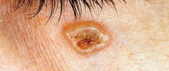
Keratosis of the skin.
Photos of adults show what neoplasms may look like. The follicular form most often develops in women. It is characterized by the formation of flesh-colored keratinized plugs on the hair follicles. New growths are uneven. Their structure resembles flat, silvery scales. The size of the neoplasms is no more than 1.5 cm.
Most often localized in the area:
- faces;
- buttocks;
- neck;
- armpits.
Keratosis of the skin (the photo in adults shows only external symptoms) begins in the cold season. In warm weather, the symptoms are erased. At a late stage of the disease, the nodules increase in size. The surface of the skin becomes rough. At the last stage, they may begin to bleed and hurt. The seborrheic appearance is characterized by slow development and chronic course.
A spot on the skin of a yellowish tint reaches a diameter of 3 cm. It has a dense morphology with scabs. At the second stage, the spot begins to increase in size and many layers form in it. The neoplasm becomes crusty. The thickness of the stain reaches 1.5 cm. On the last steel, the seal begins to turn black, crack and bleed.
The pathology is localized in the area:
- backs;
- breasts;
- faces;
- neck;
- scalp.
This type of pathology does not develop into an oncological form. In the actinic form, which often develops into a cancerous tumor, nerve rough spots appear in the first stage. At the last stage, they transform into keratinized scales of burgundy color.
Keratomas are located singly. They grow very slowly, reaching 2 centimeters. This form can disappear on its own, and appear under mechanical stress or injury.
In the initial stages of solar keratosis, a large number of small spots appear that begin to peel off. They rise above the epidermis. At the last stage of the disease, the spots transform into plaques covered with hard scales.
This form of pathology is localized in the area:
- faces;
- backs;
- soles;
- legs;
- hands
This form of the disease is considered precancerous. It is triggered by radiation. The initial stage of cutaneous horn is characterized by the appearance of gray or brown spots on the skin. At the second stage of keratosis of the skin, the spots begin to become covered with keratinized scales.
Externally, in the photo of adults, they resemble a tubercle that rises greatly above the skin. Some neoplasms take the form of a flat, silvery plaque. At the last stage, keratomas begin to peel off and bleed.
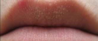
The pathology is localized in the area:
- lips;
- nose;
- century;
- forehead;
- mucous membranes of the genital organs;
- ears;
- scalp.
This form can turn into an oncological form.
Causes of keratosis of the skin
An acquired disease may appear:
- as a consequence of functional disorders of the nervous and endocrine systems;
- oncological diseases of internal organs;
- due to negative contacts with mechanical, chemical and physical effects (occupational keratosis);
- as a consequence of a sexually transmitted disease complicated by a deficiency of vitamins A, E, C;
- excessive exposure to the sun.
When the epidermal layer is damaged, actinic or senile keratosis appears.
Signs of actinic keratosis appear in a person around the age of 50. And by the age of 80, everyone has signs of this disease. This is due to the fact that the effect of ultraviolet radiation on the human body accumulates over time.
Manifestations of early disease are observed in people with fair skin, red or blond hair, and blue, gray or green eyes.
People whose bodies are less able to combat the negative effects of radiation, those with weakened immune defenses, those who have undergone chemotherapy or exposure to x-rays, and others are more susceptible to the disease.
Hereditary causes of the disease are associated with a genetic factor. If your relatives have symptoms of keratosis, then you may also develop keratomas, pilaris and other signs of the disease.
Symptoms of keratosis
Symptoms of the cyanide form include:
- the presence of multiple formations of brown or black color;
- the formation of round-shaped plaques with pronounced boundaries;
- the appearance of loose scales;
- the presence of small elevations above the level of the skin;
- formation of round or oval spots;
- formation of tumors on the back, chest and forearms;
- the appearance of plaques with a diameter of 1 mm to 2 cm.
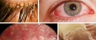
Symptoms of angiokeratoma include:
- the presence of red or dark burgundy nodules with a diameter of 1-5 mm, rarely 1 cm;
- blurred boundaries of nodules;
- irregular shape of nodules;
- arrangement of spots in groups;
- elevation of tumors above the skin level;
- expansion of capillaries.
Follicular keratoma is characterized by:
- dry dermis;
- colored nodules gray or pink;
- clear boundaries of the nodule, the diameter of which is 1.5 cm;
- tuberosity of neoplasms.
The distinctive signs of seborrheic keratoma are:
- oval or round shape of neoplasms;
- slight elevation of tumors above the skin level;
- spot diameter up to 6 cm;
- yellow or dark brown spots;
- group arrangement of spots;
- itching;
- peeling.
The actinic form, whose texture resembles sandpaper, is distinguished by:
- absence of itching and peeling;
- rashes in the form of seborrheic warts;
- plaque diameter up to 4 cm;
- bleeding and painful growths.
The main signs of solar keratosis include:
- the presence of formations in the form of groups;
- plaque formation;
- slight elevation of plaques above the skin;
- the appearance of rough scales that are easily separated.
The cutaneous horn is characterized by:
- the appearance of gray or brown spots;
- compaction of neoplasms;
- keratinization of spots;
- peeling of tissue;
- bleeding.
Symptoms
Provoking factors in the development of clinical manifestations may be:
- avitaminosis;
- stress;
- use of oral contraceptives;
- unhealthy food;
- lack of personal hygiene;
- cold weather.
General symptoms:
- pigmented skin;
- keratinization;
- the appearance of various formations or thickenings;
- itching;
- possible pain syndrome;
- bleeding
The nature of skin formations and symptoms vary depending on the form of the disease and location.
With palmoplantar keratosis, keratinization, deep cracks, and blisters appear on the heels. Roughness of the skin on the hands and feet occurs in 100% of cases, a change in the shade of the dermis from amber to brown - in 90%, increased sweating of the soles and palms - in 70%, painful cracks - in 60% of cases. Keratosis of the feet can occur when wearing uncomfortable shoes or an unsuccessful pedicure.
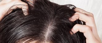
The first symptom of keratosis of the scalp is dry skin and hair, deterioration of hair condition (brittleness, dullness, split ends), dandruff. Next, keratinization appears in the area of the hair follicles, irregularities and tubercles are felt on the head, which can bleed if damaged.
As the disease progresses, hair begins to fall out in individual areas or over the entire surface at once. At the site of baldness, hair does not grow back, which is due to the death of the hair follicles.
The seborrheic form is characterized by the appearance of hyperpigmented spots, which transform into warts or plaques with clearly defined boundaries, covered with crusts. Plaques can occur anywhere on the body, plural or singular. The color of the formation varies, from flesh-colored to black. When subjected to mechanical damage (rubbing, squeezing), warts bleed.
Symptoms of the follicular form usually appear at an early age. The first manifestation is dry dermis. Next, nodules with hairs on top appear, which are located symmetrically on the skin. A peculiarity in children is keratosis on the face, usually without any unpleasant sensations other than aesthetic ones. Often these manifestations disappear during adolescence due to hormonal changes.
Skin damage in the solar (actinic) form of the pathology occurs in open areas with direct exposure to ultraviolet radiation. At risk are older people, those who spend a long time in the sun, people with fair skin, HIV-infected people, and those with age spots.
The senile form is characterized by the formation of multiple fused hard scales on open areas of the body. Round or oval lesions up to one centimeter in size are difficult and painful to remove. At risk are men, people with skin photosensitivity types I – III, who are exposed to ultraviolet radiation for a long time.
Seborrheic keratosis
Causes of keratosis
The causes of senile keratoma include:
- genetic predisposition;
- malfunction of epithelial cells;
- the presence of chronic pathologies of internal organs;
- accumulation of toxins in the body;
- violation of metabolic processes;
- degeneration of the skin.
The causes of angiokeratoma are still unclear. Some forms have a genetic basis. Most patients are diagnosed with metabolic disorders.
Provocateurs include:
- mechanical damage;
- frostbite;
- burn;
- presence of calluses;
- injuries;
- insect bites.
The follicular form can develop under the influence of the following factors:
- lack of vitamins A, C, D and group B;
- abuse of fatty, hot, spicy and salty foods;
- frequent consumption of coffee;
- use of hormonal-based drugs;
- stress and emotional stress;
- presence of syphilis;
- exposure to cold;
- HIV infection;
- tuberculosis of the skin;
- fungal infection of the feet;
- damage to the body by systemic lupus erythematosus;
- presence of scleroderma;
- allergy;
- hormone imbalance;
- prolonged exposure to x-rays;
- exposure to chemicals and poisons;
- wearing synthetic clothing;
- dysfunction of the digestive organs.
Experts have found that the pathology may have a genetic basis. The disease among adolescents and children is diagnosed in 50-80% of cases. Adult patients account for 40%.
The causes of seborrheic form include:
- heredity;
- malfunction of the immune system;
- skin aging;
- endocrine disorders;
- hormone imbalance.
Actinic form provocateurs include:
- living in a hot climate;
- presence of blond hair and freckles;
- presence of blue or green eyes;
- frequent sun burns;
- age above 45 years.
Keratosis of the skin (photos of adults indicate that this is a serious skin pathology) of the actinic variety, unlike the solar form, develops very slowly due to constant exposure to sunlight.
Causes of solar keratosis include:
- living in southern regions with a hot climate;
- exposure to stress;
- use of hormonal drugs;
- presence of blue eyes and blond hair;
- frequent sunburns.
The development of a cutaneous horn provokes:
- exposure to ultraviolet rays;
- endocrine system disorders;
- viral infection;
- presence of oncological tumors;
- skin trauma;
- genetic basis;
- obesity;
- lack of vitamins;
- malfunction of the gastrointestinal tract;
- disorders at the cellular level;
- alcohol and drug use;
- stress;
- burns;
- exposure to sunlight;
- poor hygiene.
Treatment
Treatment of hyperkeratosis depends on the causes of the disease and the form it has taken. For example, today there is nothing to treat the follicular form of the disease. If the cause of the disease is damage to internal organs, treatment of the underlying disease leads to the disappearance of the symptoms of hyperkeratosis. Symptomatic therapy is an ointment for hyperkeratosis containing fruit acids or based on lactic acid products.
Neither scrubs nor peelings can be used! This causes a deterioration in the condition and the addition of a bacterial infection (the development of pyoderma).
Abroad, treatment of foot hyperkeratosis is usually carried out in the office of a specialist doctor (podiatrist or podiatrist). In addition to treatment, the doctor recommends correcting the causes that led to the pathology:
- selects shoes;
- recommends treatment of fungal infections;
- and special individual orthopedic insoles.
There are few such centers in our country. Areas of excess keratinization are removed using special medical instruments and devices (mills, scalpels). Patients are recommended special correctors and even prostheses that help distribute the load on the feet differently.
At home, it is recommended to regularly remove keratinization using:
- pumice;
- special brushes;
- hard washcloths;
- blades;
- peelings and scrubs;
- salt baths;
- local urea-based products (Foretal Plus).
There are also special external remedies for eliminating hyperkeratoses - keratolytic creams, and moisturizing balms, pastes and gels containing urea, salicylic or acetic acid.
In the podologist's office, aggressive agents can be used - alkalis, laser and cryocoagulation, curettage.
Treatment of hyperkeratosis is long-term; if the cause cannot be found out or eliminated, therapy will be lifelong.
In severe cases, hormonal ointments are prescribed to facilitate exfoliation of the stratum corneum and accelerate healing, clobetasol or fluacinolone ointments.
Cervical hyperkeratosis is treated mainly using hardware and surgical methods:
- exposure to ultrasound;
- cold therapy (cryotherapy);
- laser correction;
- conization (electro- and using a scalpel) - removal of altered tissue;
- amputation of the uterus and cervix.
Probiotics are prescribed orally and vaginally to maintain the normal balance of vaginal microflora. Vitamin therapy (A, B 9 and C).
Diagnosis of keratosis
Keratosis of the skin (photos in adults indicate the stage of development of the disease) is diagnosed as a result of a medical visual examination. If the lesion has become large, bleeds and thickens, then there is a possibility that the neoplasm has degenerated into a malignant form.
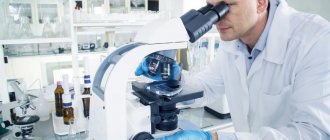
To exclude a dangerous pathology, a biopsy is performed. The procedure involves taking skin samples. It is performed by a therapist using local anesthesia. The sample taken is sent for microscopic analysis.
Also, to diagnose keratosis, dermatologists carry out:
- dermatoscopy (the procedure is carried out using a dermascope apparatus that can detect morphological changes in the skin;
- Ultrasound of neoplasms.
The most common methods are biopsy and dermatoscopy. The cost of dermatoscopy in Moscow ranges from 500 to 3000 rubles. The cost of a biopsy is 1,480 rubles. The cost of dermatoscopy in Samara ranges from 250 to 2500 rubles. The price for a biopsy is 1,500 rubles.
Diagnostics
A dermatologist can make an accurate diagnosis. But sometimes you can’t do without consulting an infectious disease specialist, venereologist, oncologist and other specialists.
The doctor will examine and palpate the affected areas of the skin. If a malignant neoplasm is suspected, a histological examination and biopsy of keratinized particles are prescribed.
If the doctor believes that the growth is infectious in nature, then the patient is sent for a blood and urine test, and, if necessary, a smear from the urethra for men and from the vagina for women.
Prevention of keratosis
In order to avoid complications, you should adhere to the following preventive rules:
- use creams with a moisturizing effect for the skin;
- include more foods containing vitamins in your diet;
- limit exposure to sunlight;
- wear special clothing and gloves when working with chemicals;
- do not sunbathe in the sun or in a solarium;
- strengthen the immune system;
- avoid skin injuries;
- maintain hygiene;
- When diagnosing dermatosis, undergo regular examination by a dermatologist.
Symptoms of manifestation
The initial symptoms of keratosis are not always noticeable. Their manifestations in the form of small irregularities and roughness can be found on the cheeks, ears, bridge of the nose, neck - in those places that are exposed to the sun.
- keratinization of hair follicles;
- lumpiness of the skin surface;
- peeling of the skin;
- bleeding of cracks;
- erosive manifestations;
- the appearance of painful sensations.
It is necessary to seek advice from specialists if:
- the tumor grows rapidly;
- the bleeding of changes does not stop;
- the color of the tumor changes, pain and itching stop.
Treatment methods for keratosis
Keratosis is eliminated through drug treatment. Folk remedies are used as additional therapy and only after consultation with a specialist. Physiotherapeutic procedures are used to relieve pathology. In case of complicated course of the disease, they resort to the surgical method.
Medications
| Antibiotics | Zinnat, Doxycycline, Azithromycin |
| Ointments | Efudex, Fluorouracil, Imicvod, Fluoroplex, Solcoderm |
| Vitamin complexes | Milgamma, Bnevron BF, Vitamin A Bartel Drugs, Retinol |
The drug Zinnat is used for the inflammatory process. Prescribed 500 mg 2 times a day. The course of treatment is 7-10 days. The cost of the drug 500 mg is 420 rubles. There are 10 tablets in a package.
Imicvod ointment is used 3 times a week. Treatment should continue until symptoms disappear completely. The maximum course of treatment is 4 months. The price of the drug 5% is 5087 rubles. The tube contains 250 mg of the substance.
Vitamin A capsules Bartel Drugs take 1 tablet per day 15 minutes after meals. The duration of treatment is determined by the doctor. The cost of the drug is 150 rubles. There are 50 capsules in a package.
Traditional methods
To relieve the symptoms of keratosis, you can use folk remedies:
- Agave leaves, after three days of freezing, are applied to the affected area overnight. After removing the compress, the keratome is treated with salicylic alcohol. The course of treatment lasts 3 weeks.
- Grated raw potatoes are applied to the affected area. The top of the potatoes is covered with a clean cloth and cellophane. After 40 minutes, the bandage is removed and the potatoes are washed off with water.
- Take 2 tbsp. l. dry leaves of celandine. They are filled with 250 ml of boiling water. The mixture is infused for several hours. Lotions are made on its basis. You can wipe the skin with the infusion.
- Take 1 tbsp. l. onion peels. Pour 1 tbsp of vinegar into it. Leave the mixture in a dark place for a week, then strain. It is recommended to make lotions. They are left on the affected area for 30 minutes. It is important to avoid contact with healthy areas to avoid burns.
Other methods
Since conservative treatment is not effective in all cases, they resort to drastic measures - removal of growths. They are especially used when there is a possibility that the tumor will degenerate into a cancerous tumor.
The following types of treatment are used:
- Cryodestruction . It involves freezing the growths using liquid nitrogen. The method is used when the skin lesion is extensive. When frozen, plaques undergo necrosis. A possible complication of the operation may be increased skin pigmentation.
- Removal via radio waves . This manipulation involves excision of the tumor with a radio knife under the influence of radio waves.
- Electrocoagulation. Represents cauterization using high-frequency electric current. Performance involves anesthesia. There is a risk of scarring and age spots.
- Laser destruction . A targeted effect on the keratoma with a carbon dioxide laser is used. This is the most effective method of treating keratosis, which is characterized by painlessness, speed and absence of side effects.
- Photodynamic therapy involves the application of methyl aminolevulinate followed by exposure to a light wave of a given length. This process causes tissue necrosis. In this case, healthy tissues are not damaged. After the procedure, you should not be in the sun for 48 hours.
- Curettage is used to treat solar keratosis . This is an operative method in which individual areas of the lesion are scraped out using a curette. Curettage is not used to treat the actinic form, as it can cause scarring.
- Dermabrasion . Removing tumors using an abrasive brush.
- Plasmolifting involves the use of plasma injection, which stimulates local immunity and promotes skin regeneration.
- Biorevitalization involves injections of hyaluronic acid, which improve the morphology of the skin, promote rejuvenation, and increase the level of regeneration.
- Chemical peeling . It is used for minor lesions. For chemical treatment of warts, lactic or glycolic-salicylic acid is used.
Treatment of keratoma
Most types of keratosis do not pose a threat to human life, bringing only aesthetic discomfort. At the same time, spots on the skin are susceptible to transformation into malignant ones, and there is also a risk of damaging them due to friction or pressure from clothing, or accidentally touching them, which will lead to inflammation and infection. Injured areas of skin can become infected with the human papilloma virus or herpes.
Before treatment, the dermatologist evaluates what skin keratinization looks like, what caused it, and determines the type of keratosis. Conservative methods of getting rid of tumors include:
- removal by laser, electrocoagulation, excision under local anesthesia;
- precancerous tumors are removed by administering cytostatics and antibiotics;
- applying ointments to reduce the size and healing of keratoma on the skin;
- introduction of special preparations (Solcoderm) for resorption of seborrheic keratosis.
Here is a brief treatment plan for keratosis depending on the type of pathology:
| Type of keratosis | Keratoma Treatment Options |
| Seborrheic | Laser, nitrogen, prospiridone or fluorouracil ointment |
| Senile | Laser, electrocoagulation, surgical removal |
| Solar | Use of liquid nitrogen, laser, antitumor ointments |
| Follicular | Electrocoagulation, scalpel |
| Cutaneous horn | Surgical removal, laser, radio wave exposure |
Treatment of seborrheic keratosis at home
In addition to conservative therapy, seborrheic keratosis can be treated at home:
- lotions from thin plates of aloe leaves at night, wiping with salicylic alcohol in the morning;
- applying a piece of propolis for two weeks;
- lotions made from an infusion of onion peels in vinegar.
Treatment of actinic keratosis
If the growths are safe and without bleeding, treatment of actinic keratosis can be carried out at home:
- lotions of warm castor or sea buckthorn oil;
- garlic gruel with honey;
- gruel from raw potatoes.
Treatment of actinic keratoses
It will not be possible to completely get rid of tumors at home, but treatment of actinic keratosis can be reduced to softening the crusts and reducing the size of the spots. For this use:
- compresses made from warm vegetable oils;
- rubbing walnut fruits with infusion;
- lotions made from a mixture of celandine leaves and pork fat.
Removal of cutaneous horn
Only surgical removal of the skin horn is carried out, because the formation is too large and will not disappear at home. A pronounced cosmetic defect is subject to surgery even if there are no results from other treatments. Removal of keratomas occurs in the following ways:
- laser - exposure to a neodymium or erbium laser beam, the layers of the spot are dried, leaving a small scar, the procedure lasts 15 minutes;
- freezing with liquid nitrogen – used to eliminate single keratomas;
- electric knife – small keratomas are removed, excision is performed with a minimal percentage of trauma;
- radiosurgery is a modern method that does not leave scars on the skin;
- a scalpel is a traditional way to remove large tumors and adjacent affected tissue.
Ointment for keratosis of the skin
To soften the keratinized areas, use ointment for keratosis of the skin. Apply it in the form of applications according to the instructions. Keratoma on the face is treated with the following drugs:
- Ureatop, Keratosan, Ureaderm, Akerat - keratolytics with urea in a concentration of 12-30%;
- Efudex, Fluoroplex, Carac – contain fluorouracil;
- Aldara – contains imiquimod;
- 3% Diclofenac gel in small doses.
Possible complications
The main complications of keratosis include:
- development of microbial eczema;
- dysfunction of the nervous system;
- degeneration of growths into a cancerous tumor;
- tooth loss.
Keratosis of the skin is fraught with serious complications, and the photo of adults shows that this pathology is a serious skin disease that requires treatment by a dermatologist and excludes self-medication.
Article design: Oleg Lozinsky
Causes
Various negative factors contribute to the development of keratosis:
- decrease defenses ;
- chemical effects on the skin;
- heredity;
- history of infectious
- changes in the condition of the epidermis that occur with age.
The following groups of people are susceptible to the disease:
- carriers of HIV and AIDS;
- those who have undergone complex operations;
- patients with disorders of the endocrine system;
- suffering from diseases of the nervous system;
- patients who have undergone chemotherapy.
In some cases, the reasons for the appearance of compactions remain unclear.
Prevention of hyperkeratosis
Compliance with the rules of prevention will avoid both the onset of the disease and remissions. To prevent keratoses it is important:
- limit your intake of foods high in saturated fats and refined foods;
- eat vegetables and fruits high in vitamins A, C, E;
- follow the rules care not only for the face, but also for all areas of the body;
- select clothes and shoes according to size;
- use high-quality cosmetics to combat excessive dry skin;
- get rid of excess weight;
- avoid stress, keep the nervous system in order;
- cure diseases of internal organs in a timely manner.
It is known that the condition of the skin shows the level of human health.
Diagnostic methods
Only an experienced dermatologist should treat keratosis of the skin. If the slightest symptoms of the disease appear, it is important to consult a doctor as soon as possible.
Keratosis takes a long time to cure; in addition, the patient must follow a diet rich in fats and vitamins. You can also apply a medicinal ointment prescribed by a dermatologist to the tumor yourself at home.
The treatment method is selected individually for each patient, taking into account his tests, characteristics of the disease, and the general condition of the patient. But before this, the dermatologist must decide how to more effectively deal with the type of keratosis, because everyone’s disease progresses differently.
It is important to diagnose the disease correctly, and also to find out its complexity, type and location. There are three diagnostic methods:
- Biopsy (removal of material for histological examination).
- Histology (study of tissues of the human body).
- Standard inspection of covers.
Only after diagnosis will the dermatologist select the individual and correct treatment for each patient.
Prevention measures
Preventive measures include:
- Avoid going to the solarium.
- Immunity support, additional intake of all necessary vitamins.
- Proper nutrition.
- Protection from chemicals (usually entering the body through household detergents).
- Avoiding ultraviolet rays.
- Use of sunscreens.
For people whose work activity involves the street (especially under the scorching sun in the summer season), it is important not to forget about skin protection. Before work, apply sunscreen every day (protection level above 30). If possible, it is better to go into the shadows.
Additionally (especially in winter) drink vitamins and tea with lemon. It is better to wear protective gloves when washing dishes and cleaning the apartment.
The body needs constant monitoring and protection. Leather also requires care and additional protection. Some neoplasms seem insignificant; they do not bother, do not itch or increase in size. But a benign tumor can always develop into cancer.
It is better to consult a doctor in a timely manner and get rid of keratosis. Otherwise, in advanced cases, drug treatment will not correct the situation, and the intervention of an oncological surgeon will be required.
Skin hyperkeratosis - what is it?
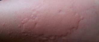
When areas of keratinization develop on the skin, the clinical presentation may vary between conditions. Common features of all forms of skin hyperkeratosis are excessive dryness of the skin, coarsening and lumpiness.
The affected areas lose sensitivity, but when pressing on them (callus), pain is possible due to irritation of the nerve endings by the rough tissues.
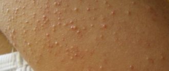
Follicular hyperkeratosis (photo)
The skin on your elbows, knees, outer thighs, and buttock folds is naturally drier. The more keratinized scales are formed by the integument of the body, the more difficult it is for them to be rejected and the easier it is for them to accumulate.
Dead cells in follicular hyperkeratosis cause blockage of hair follicles. Clogging into their mouths, epithelial scales cause inflammatory changes there, leading to the formation of dense tubercles.
If you run your hand over areas of the body affected by keratosis pilaris, a sensation of goose bumps will appear. Dense bumps may be barely visible or appear as bright red or brown spots.
Accidental injury or attempts at squeezing often lead to microbial infection of the follicles, their suppuration, and the development of complications - pyoderma.
Hyperkeratosis of the foot
Of all the parts of the body, it is the foot that bears the maximum load. It accounts for the entire body weight. It is easy to understand that with obesity, static and dynamic effects increase many times over. In addition, long walking and “standing” work, when a person has no time to rest, have a negative impact.
On the other hand, people rarely think about their feet. Dress shoes with high heels, lack of proper correction of flat feet and other pathologies only aggravate the situation.
Compensatoryly, the body seeks to protect itself. In the epidermis of the feet, in response to adverse effects, horn cells begin to actively form, their number increases, the skin thickens, becomes insensitive, and rough. If a woman constantly wears high-heeled shoes, the maximum load on the forefoot causes keratinization in this particular area.
Standing work is the key to hyperkeratosis on the heels. With flat feet, the middle part of the foot (arch) can become rough. If a limited area is exposed to excessive stress or systematic friction or pressure, a callus will form.
Hyperkeratosis of the foot photo of cracks
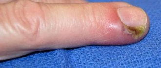
In addition, hyperkeratosis always develops with a fungal infection. If the causative agent of mycosis settles on the nail, subungual hyperkeratosis develops. This condition is characterized by an increase in the number of keratinocytes in the skin. As a result, clusters of these cells form, raising the body of the nail, which looks like its thickening.
A common occurrence is keratinization of the skin of the feet in diabetics . It is caused by tissue trophic disorders. It is this category of people who should monitor foot hygiene more carefully than others, carefully select shoes of the appropriate size and high quality, and also use various foot correctors.
A fairly common complication of advanced foot hyperkeratosis is the formation of cracks. It is caused by a loss of skin elasticity combined with the accumulation of an impressive mass of dead skin cells.
- Such cracks are impressively deep and difficult to treat.
Hyperkeratosis on the face - what is it?
“Hyperkeratosis on the face - what is it and how to treat it?” - Frequent questions with which patients turn to their doctors. In this case, the age category does not matter, since the disorder occurs at any age. In scientific terms, the pathology is characterized by keratinization of tissue due to the formation in certain layers of cells of a substance consisting of keratohyalin, keratin and fatty acids. The combination of these elements causes growths that cause discomfort, and symptoms depend on the severity of the disorder.