Basal cell carcinoma or skin basal cell carcinoma occupies 1st place in the structure of oncological diseases. As a rule, it affects both men and older women equally. But recently there has been a tendency towards “rejuvenation” of the disease. Young patients are increasingly turning to oncologists. This is certainly due to uncontrolled visits to solariums, excessive sun exposure and untimely visits from patients with precancerous skin diseases.
Make an appointment
Basal cell skin cancer is formed from the basal layer of the skin, is usually localized in one place and does not metastasize. But there is multiple basal cell carcinoma - this is when one patient simultaneously has several foci of tumors located in different areas of the skin. the number of such foci can range from two to 20 or more. Skin basalioma does not metastasize, but if not treated in a timely manner, it can destroy not only all layers of the skin, but also cartilage and bones, thereby causing patients not only aesthetic defects, but also physical suffering. Skin basal cell carcinoma has a tendency to degenerate into metatypical and squamous cell skin cancers, which metastasize to lymph nodes and other organs. The size of basalioma varies from 0.5 cm to 20 cm or more. As a rule, basal cell carcinoma is localized on the face in 80% of cases, which complicates treatment.
Organs such as the auricle, wing and slope of the nose, the corner of the eye and the periorbital area, the red border of the lip, and the scalp are inconvenient locations and not always traditional treatment methods such as surgery or close-focus radiotherapy can bring success and patient satisfaction with the treatment. After all, the consequence of surgical treatment can be aesthetic defects: scars, deformation of organs, and even their loss. Close-focus X-ray therapy also has consequences, such as chronic perichondritis, chronic sinusitis and others, since along with neoplasms it also affects healthy skin tissue, as well as nearby tissues (mucous membrane, cartilage and bones).
How to cure basalioma?
Basalioma is a neoplasm, one of the types of skin cancer, which is classified as neither malignant nor benign tumors. Treatment for basal cell carcinoma usually involves its removal. However, it is necessary to take into account the characteristics and stage of the disease.
Basalioma: what is it?
Basal cell carcinoma is called basal cell carcinoma of the skin. Despite ongoing research, it is still unknown from which cells the tumor is formed.
It rarely metastasizes and grows slowly. If treatment for basal cell skin cancer is started promptly, complications can be avoided. This type of skin cancer mainly occurs in people over 50 years of age, regardless of gender.
Basalioma can manifest itself in different ways.
There are the following types of tumor:
- Superficial. Occurs more often than others. It looks like a small red spot on the skin, which then begins to itch and peel.
- Nodular. In the initial stages it resembles a dense pimple. Over time, it grows, the skin in the area of the nodule becomes thinner, and blood vessels become visible through it.
- Ulcerative. It usually starts as a nodule, but then an ulcer appears in the center of the formation, which can fester.
- "Turban" The tumor affects the scalp. Several pink nodules form.
- Nodal. It usually affects the face and neck. It first appears in the form of small dense nodules, which eventually merge into one tumor.
- Warty. Consists of several uneven compactions, looks like a head of cauliflower.
- Pigmented. Basalioma resembles a dark mole, but its surface is uneven and consists of tiny nodules, on the surface of which ulcers can form over time.
- Scar-atrophic. The neoplasm is an ulcer. A scar forms in its center, but along the edges the ulcer continues to grow.
These forms of tumor differ not only in appearance, they can occur in different ways. Treatment of facial basal cell carcinoma in Moscow is carried out in various ways. But it is important to start it as early as possible. Basalioma can spread deeper, destroying healthy tissue.
Why is basal cell carcinoma dangerous?
Basalioma is often characterized by a benign course. There are very few cases of metastases in medical practice.
However, this disease is a type of skin cancer, so it cannot be ignored.
Treatment of basal cell carcinoma of the face with ointments rarely gives a positive result. To avoid further spread of cancer cells, the tumor must be removed, which, unfortunately, is not always possible. Basalioma can be located in dangerous places, for example, close to the eye.
The danger of basal cell carcinoma is that it grows into the deep layers of the skin, especially some of its forms.
The tumor affects other tissues and eats them away. If it is located close to the eye, cancer cells affect the organ of vision, which leads to blindness. Facial muscles, nerves, cartilage and even bones can suffer from the growth of tumor tissue.
If the disease is aggressive and quickly penetrates deep into the tissue, more serious consequences are possible, including damage to brain tissue. “Turban” basal cell carcinomas are especially dangerous in this regard.
For safety and to prevent serious consequences, it is better to remove any manifestation of basal cell carcinoma.
If the tumor is located on the body, it is less dangerous, but still requires surgery. However, basaliomas on the body are not so common; they usually affect open areas of the body.
Causes of basal cell carcinoma
Since basal cell carcinoma is a skin cancer, it is difficult to determine the causes of its appearance. It is believed that this disease can be inherited. The likelihood of encountering basal cell carcinoma if your close relatives have it increases several times.
Experts have also identified provoking factors:
- Exposure to ultraviolet radiation;
- Ingestion of carcinogens into the body;
- Genetic predisposition;
- Injury to moles;
- Bad habits;
- Congenital skin pathologies;
- Skin diseases and viral infections.
photos on the Internet after treatment for basal cell skin cancer. With timely detection and removal, unpleasant consequences and cosmetic defects can be avoided. But prevention is no less important. It is advisable to avoid provoking factors.
Particular attention is paid to sun exposure and solarium abuse.
Such an aggressive effect on the skin provokes a genetic change in the structure of the cell, which can trigger the development of the disease.
It is believed that people with thin and fair skin and freckles are more susceptible to negative factors and are more prone to skin cancer.
This skin reacts more strongly to the sun's rays.
One of the causes of basal cell carcinoma may be decreased immunity. Various infections are more difficult to tolerate. The herpes virus can also lead to skin cancer.
Smoking also has harmful effects on the skin, which absorbs carcinogens, mutagens and other substances from cigarette smoke. All this reduces the protective functions of the skin and contributes to the thinning of the walls of blood vessels.
It is important to promptly treat various skin diseases, eczema, dermatitis, and follow the rules of scar care to avoid their suppuration.
All this significantly increases the risk of encountering basal cell carcinoma. For this reason, prevention plays an important role. Doctors recommend protecting your skin from sun exposure, using sunscreen, avoiding skin injury, protecting it if you have to work with harmful substances, and promptly treating skin diseases.
Laser tumor removal technique: how it works
Basalioma is a malignant tumor. It develops when there is a failure in the process of cell division in the basal layer of the dermis. It does not have a capsule, so the process of growth of the formation occurs with the capture of tissue simultaneously in breadth and depth. Over time, damage to cartilage and bone tissue becomes possible. If basal cell carcinoma forms on the face, close to the eyes or ear canal, or on the head, the patient may lose hearing and vision. When the brain is damaged, the patient dies.
The tumor does not resolve on its own. It grows slowly but constantly and increases by 5 mm per year. The part that forms on top of the skin looks like a mole or wart. The invisible region is similar to a cone or ellipse.
To prevent life-threatening complications, the tumor is removed. Recently, a laser has been used to implement this, an installation that reproduces a directed beam of light of a certain wavelength. The surgeon has the ability to control the depth of penetration of the laser, which passes through a complex system of mirrors and is fed through a special nozzle. It can be used to make micro-incisions. Heat irradiation causes water molecules to move faster. As a result, the liquid located in the cells of the tumor focus evaporates. Dehydrated, they begin to die.
In Russia, a CO2 laser is used to carry out such operations. The Lancet installation reproduces it in continuous mode. It helps to perform vaporization (evaporation of pathological tissue layer by layer) without damaging healthy cells. In parallel with this, coagulation of blood vessels occurs. It prevents bleeding. Nerve endings are also cauterized, so in the postoperative period the patient does not experience severe pain. Thermal irradiation disinfects the surgical field and minimizes the risk of infection of the resulting wound.
Radiation treatment of basalioma.
On the one hand, radiation is quite effective, giving an average of 8.7% relapses. On the other hand, treatment of basal cell carcinoma is quite lengthy, requiring several sessions over 1 month. In addition, relapses after radiation therapy are the most dangerous, since the rays are radioactive and damage the DNA of skin cells. This is also associated with a high risk of the appearance of new basal cell carcinomas and squamous cell skin cancer near the irradiated area. We also remember that patients already have a high risk of developing new basal cell carcinomas. Patients under 65 years of age have a longer life expectancy and, accordingly, a greater risk of developing radiation-induced skin cancer. In large Western countries, radiation treatment is contraindicated for people under 65 years of age. There is also a risk of complications, such as radiation necrosis and scalp hair loss. Treatment of basal cell carcinoma by irradiation more >>>
Hair loss and multiple new tumors that appeared many years after radiation treatment for basal cell carcinoma.
Chronic radiation necrosis (radiation) several years after radiation treatment of basal cell carcinoma. As a result of DNA damage, the skin cells did not turn into a tumor, but began to function much worse.
Advantages and disadvantages of the technique
Using a laser, you can remove tumors located in hard-to-reach places. The operation lasts 15-20 minutes. After it, a black crust forms on the wound. It creates additional protection for the penetration of pathogenic bacteria. Natural skin healing processes take place underneath it. Therefore, if you follow the doctor’s recommendations, you can count on a good cosmetic effect: after the crust falls off, a section of new epidermis forms at the site of the wound. It has a bright pink color. Over time, it fades and the hole becomes less noticeable. The absence of scars is another undeniable advantage of laser therapy.
The patient is discharged home on the day of surgery. With proper care of the wound area, the rehabilitation process lasts a week. This is half the cost of traditional surgery.
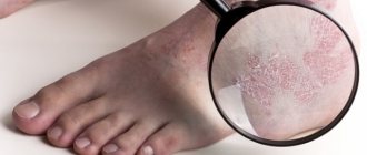
How does the disease manifest itself?
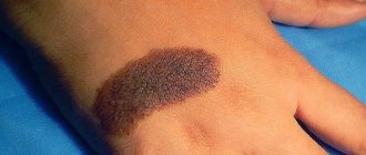
The development of the tumor begins with the appearance of a pale pink nodule on the face (skin of the eyelid, lip, in the area of nasolabial folds) and neck. Outwardly, it is very similar to a pimple, but, unlike it, it does not cause any pain. There is always a dense ridge around the formation. It becomes more noticeable if you try to pull back the skin around the nodule.
Schematic representation of basal cell carcinoma
The structure of the roller is formed from small granular elements, shaped like a pearl. Over time, a grayish crust appears in the center of the formation. If it is removed, a dimple remains in its place. Further growth of basal cell carcinoma can occur in different ways: it all depends on what type of tumor develops.
Indications and contraindications
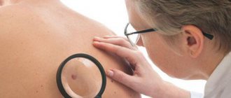
Basalioma is difficult to diagnose by appearance. It is also impossible to identify the formation using a blood test for tumor markers. In order to be able to make an accurate diagnosis, the doctor must write the patient a referral for an MRI or CT scan, perform a biopsy of the affected area and send biological material for microscopy and histology. The results of the examination make it possible to confirm the development of basal cell carcinoma, determine its exact size, and the features of germination into neighboring tissues.
Indications for laser tumor removal:
- education is in the first stage of its development;
- the lesion does not affect cartilage and bone tissue;
- the tumor constantly changes color;
- basal cell carcinoma becomes covered with a poorly healing ulcer;
- the formation is constantly exposed to mechanical trauma.
The surgeon may decide to perform laser removal if the patient relapses after using another treatment method (for example, cryodestruction or electrocoagulation).
Indications for which laser removal of basal cell carcinoma is contraindicated:
- diabetes;
- acute inflammatory processes;
- diseases of the cardiovascular system;
- poor blood clotting.
In women, surgery cannot be performed during pregnancy, lactation, or during menstruation.
Diagnostics
When a person comes to the hospital, diagnosing skin cancer begins with a visual examination. Next, the doctor takes fingerprint smears or scrapings from the surface of the tumor and refers the patient for cytological and histological examination. If nest-like clusters of spindle-shaped, oval or round cells are present, then to confirm the diagnosis it is necessary to determine the cellular composition, for which a scraping is taken from the bottom of the ulcer.
With the help of analysis for tumor markers, cancer can be diagnosed more quickly, however, with malignant basal cell carcinoma, specific tumor markers are not produced. Therefore, in all cases, a laboratory blood test is performed, during which an increased level of leukocytes, an increased ESR (erythrocyte sedimentation rate), a positive thymol test, and an increase in C-reactive protein are noted.
Since these laboratory indicators can be present in many inflammations, and the histological and clinical picture of the tumor can be varied, it is necessary to carry out differential diagnosis to help exclude or confirm other skin diseases, such as lupus erythematosus, lichen planus, seborrheic keratosis.
We recommend reading Follicular tumor of the thyroid gland - causes, symptoms, treatment
An inexperienced doctor may confuse flat superficial basalioma with Bowen's disease, the pigmented form with melanoma (cancer of a mole), sclerodermal tumor with scleroderma and psoriasis. Due to the fact that basal cell carcinoma may have more common symptoms than other tumors, the examination must be carried out very carefully and only in a good diagnostic center.
Preparation technique and process of removal of formation
There is no special preparation for laser treatment of basal cell carcinoma. After the tests are completed and deciphered, a date for the procedure is set. The operation is performed on an outpatient basis. A sick leave certificate is not issued to the patient.
The patient is asked to take a position convenient for the necessary manipulations and local anesthesia is given. The hair is covered with a special cloth to prevent inflammation. When basal cell carcinoma is removed from the eyelid, protective glasses are put on the patient's eyes.
If the procedure is performed without the use of sensitizing agents, the surgeon pre-treats the surgical field with an antiseptic solution and begins excision of the basal cell carcinoma with a laser. He does this using a special attachment, which helps the doctor fix the beam on the tumor. In essence, the surgeon has a high-precision electric knife in his hands, with which he cuts off the formation layer by layer and at the same time seals the vessels. There is no contact between the skin and the instrument, so the risk of infection is minimal. During the operation, the surgeon removes the tumor and captures five millimeters of healthy tissue (which may contain cancer cells).
There are basaliomas, the cells of which demonstrate high resistance to the effects of a light beam. In this case, laser treatment is combined with photodynamic therapy. Before the operation begins, the patient is given injections, solutions of which increase the sensitivity of the skin to light exposure. After 2-3 hours, sensitizing agents penetrate into the tumor cells. Then laser irradiation is performed. In this case, the chances of a successful operation increase significantly.
Treatment of basal cell carcinoma. Basic goals.
Treatment of basal cell carcinoma should primarily be aimed at maximum effectiveness, expressed in fewer relapses. Relapse is the reappearance of the same tumor in approximately the same place. Why is this so important, you ask, since you can repeat the treatment if it was unsuccessful the first time? The fact is that basal cell carcinoma is still a skin cancer and has the characteristic features of cancer. After an unsuccessful attempt at treatment with any method, basal cell carcinoma cells largely adapt to it due to mutations that constantly occur in any malignant tumor. Repeated treatment of basal cell carcinoma, if it is a relapse, will be less effective, giving, for example, up to 40% of relapses, if the first gave only 10%. In addition, if treatment is ineffective, the cells can spread in depth and width, worsening the initial stage of the disease and the prognosis. Also keep in mind that within 3 years, every 3rd (35% of patients) will develop a new basal cell carcinoma in a new location (not a relapse or metastasis). Within 5 years, every second person (50% of patients) will develop a new skin basal cell carcinoma that is in no way related to the previous one. In this regard, treatment of basal cell carcinoma with methods with a stimulating immune effect, such as photodynamic therapy and cryodestruction, are becoming more popular. It reduces the likelihood of new basal cell carcinomas.
Rehabilitation after laser treatment
Since laser treatment does not have the most common postoperative risks, the patient does not need to stay in the hospital. He can carry out all care of the wound surface at home. For these purposes it is necessary:
- Wash the wound twice a day with a liquid antiseptic (a weak solution of potassium permanganate) or calendula tincture;
- Cover the top of the wound with a sterile gauze bandage and change it every time after treating the scab.
During the first month you cannot sunbathe, visit baths and saunas, or swim in open water. It is also forbidden to peel off the scab yourself: natural healing processes take place underneath it, and their disruption leads to the formation of a scar. Complete healing of the wound occurs in 20 days.
If sensitizing agents were used during the operation, rehabilitation proceeds differently. In this case, swelling often forms around the wound, the inflamed skin turns red and peels. The patient experiences pain in the first days. To eliminate them, an anti-inflammatory drug (Nimesil) may be prescribed.
In the first week after surgery, you should not go outside during the daytime, stay in a room with bright electric lights, or sit in front of the TV for a long time. The healing process of the wound lasts about a month. It is accompanied by severe itching. It can be alleviated by treating the skin with ointments that contain Digoxin.
Possible complications after the procedure
Practice shows that if you follow all the recommendations of the attending physician, there are no risks of complications: there is no postoperative bleeding, swelling, inflammation, or severe pain.
However, some patients noted a lack of sensitivity at the site of basal cell carcinoma. Doctors note that she recovers on her own after several months.
Violation of processing rules leads to infection. It manifests itself in the form of redness of the skin around the surgical field, pain and swelling. In severe advanced cases, attacks of fever occur. If such symptoms appear, you should immediately seek help from a doctor.
Classification
Based on the international classification of basal cell carcinomas, there are three main types of skin cancer:
- Fibron-epithelial;
- Scleroderma;
- Surface.
Tumors can be:
- single - the disease is characterized by one focus, which is sometimes difficult to notice, especially if it is basal cell carcinoma of the scalp;
- multiple basal cell carcinomas - diagnosed in the presence of a large number of foci of the disease.
Tumors can be located on absolutely any part of the human body: back, chest, forehead, and even affect the legs. In most cases, basalioma is localized on the skin of the face and head. When the disease is localized on the scalp, multiple and single basal cell carcinomas, the presence of which is difficult to notice under the hair, are often diagnosed in the later stages of development.
We recommend reading Tumor in the vagina: symptoms of neoplasms and treatment
The following main types of basal cell carcinoma are distinguished:
- Superficial basalioma - this form appears in the form of a plaque with a diameter of up to 3 cm. They usually have a red-brown color and a large number of small vessels. On the surface, a flat plaque has crusts that can be eroded, but despite this, the superficial form of the tumor is favorable.
- Warty (papillary) - this form appears in the form of a nodule that rises above the skin. This type of tumor is not characterized by superficial growth.
- Pigmented basal cell carcinomas - such tumors are presented in the form of a pigment containing melanin, which accounts for its dark color and similarity to another form of malignant tumors - melanoma.
- Cicatricial atrophic basal cell carcinoma (scleroderma-like) - this type looks like a scar, which is localized under the skin level. During development, the tumor may alternate between scarring and erosion, as a result of which the patient experiences fresh erosions and tumor scars. Gradually, the neoplasm grows and affects neighboring areas of the skin, and scars form in the middle.
- Ulcerative basaliomas (or nodular-ulcerative) - such tumors pose a serious threat, as they rapidly destroy the soft tissues that surround the tumor. Most often, ulcerative basaliomas are located on the corners of the eyes and on the eyelids, so the disease is often called basalioma of the eyelid or basalioma of the eye. The tumor can also occur on the scalp and in the nasolabial folds.
- Solid basalioma (nodular) - this form of the disease is located in the same areas of the skin as the previous type, but is distinguished by the growth of skin formations outward, and not deep into the skin layers. Externally, this tumor form of basalioma resembles half a ball protruding above the skin and has a yellowish or light pink color, with raised edges. Solid basal cell carcinoma of the skin of the back is most often observed, but it can also occur on other areas of the skin. Due to the dark pigmentation, when diagnosing this tumor, it must be differentiated from other tumors and other skin lesions, in particular from melanoma.
- Adenoid basalioma - cells of this form are characterized by the creation of structures similar to glands and tubes, which is why the neoplasm takes on an openwork appearance. This is a recurrent tumor, which is characterized by the presence of colloidal mucoid content in the lumens of the structures. Often occurs together with the solid form of the disease.
- Warty basal cell carcinomas are often mistaken for ordinary warts, as they appear as dense nodules growing out of the skin. The positive quality of such a neoplasm is that it does not grow into healthy tissues of the body.
Nodular forms of basalioma are diagnosed most often and are characterized by the appearance of a small red nodule on the surface of the skin. During development, the nodule may ulcerate, resulting in the formation of depressions covered with a crust on the surface.
The nodular form of basalioma is usually localized on the eyelids and facial skin. If a patient has basal cell carcinoma of the lower eyelid, it can pose a serious threat to the organs of vision, therefore, if you suspect the presence of this disease, you must be examined by a doctor. When basal cell carcinoma forms, symptoms, diagnosis and treatment methods directly depend on its type and location.
Surgical removal of basal cell carcinoma
It is possible to completely remove basal cell carcinoma with a laser if the size of the tumor lesion does not exceed 2.5 cm. The risk of relapse in this case does not exceed 5%.
When the formation grows to 5 cm, laser therapy is used in combination with chemotherapy or radiation. This treatment is carried out in an oncology clinic.
If the mutation process is extensive, the tumor grows into all layers of the dermis and invades cartilage and bone, then it can only be removed surgically. The operation is performed under local anesthesia. The patient must come to the surgical hospital on the appointed day on an empty stomach. The day before, he must adhere to a restrictive diet and refuse to eat fatty, “heavy” foods.
The surgeon, based on the results of MRI and CT, first marks the schematic boundaries of the tumor and the desired indentation. He can perform further manipulations with a scalpel or radio knife. Using a surgical instrument, he cuts out the tumor, stops the bleeding, treats the wound with antiseptic solutions and stitches it up.
Then the recovery period begins.
In the first days, the sutures are washed twice a day with soap and water, dried with a cotton swab, and then treated with erythromycin ointment or silver sulfadiazine. When symptoms of suppuration appear, instead of soap and water, Miramistin or Prontosan solutions are used to wash the sutures.
For rapid healing of the wound surface, the patient is prescribed multivitamin complexes (“Multitabs”, “Vitrum”). This is especially true for older people.
Antibiotics are prescribed to prevent infection.
The sutures are removed on the sixth day. The entire skin healing process can take 1.5 months. Until the wound heals completely, the patient is prohibited from visiting baths and saunas, swimming in open water, or sunbathing under the rays of the active sun.
In the first year after surgery, the patient must undergo preventive examinations every 3 months. They make it possible to detect a relapse at the earliest stages of development and take steps to eliminate it.
Modern methods of treating basal cell carcinomas
Treatment methods for basal cell carcinoma can eliminate the tumor and minimize the likelihood of relapse . Modern techniques make it possible to minimize the appearance of scars, thereby restoring the healthy appearance of the skin.
Laser removal
Removing a tumor using a laser is considered one of the most popular procedures . Using a specialized dermatological laser has its advantages:
- minimal bleeding;
- fast healing;
- relative painlessness of the procedure;
- low probability of infection getting into the wound;
- pronounced cosmetic effect.
However, this procedure also has certain disadvantages:
- Do not use a laser to remove tumors larger than 2 cm in diameter;
- the presence of chronic diseases and diabetes is a contraindication to laser excision;
- the procedure is not performed on pregnant women and women during lactation;
- Laser removal is not used for hypersensitivity to light.
No specialized preparation is required to perform the procedure. Before the procedure begins, the patient undergoes the necessary tests, after which he needs to appear for removal.
itself takes no more than 30-40 minutes . The impact site is disinfected and treated with an anesthetic composition based on lidocaine. To increase the effectiveness of anesthesia, injection of the drug can be used.
High temperature promotes the “evaporation” of the changed cells, and nearby blood vessels coagulate. After a few hours, a crust forms at the site of exposure.
Healing occurs within 10-20 days . During this period, it is not recommended to peel off the crust. You can protect it from drying out with Vaseline. Before contact with open sunlight, you should use a sunscreen with a high degree of protection.
The advantage of this technique is the low likelihood of complications . This technique is great for eliminating small tumors.
Cryodestruction
Destruction with liquid nitrogen has a very high efficiency (no more than 7.5% of relapses) in removing BCC. The use of cryodestruction is one of the safe and effective methods that can eliminate tumors with a minimum amount of scars.
This procedure has its advantages:
- impact on tumors of any size and in any part of the body;
- carried out without hospitalization;
- immediately after the procedure, the patient can begin his usual activities;
- performed under local anesthesia;
- the possibility of performing procedures during pregnancy and lactation;
- no blood loss;
- Possibility of carrying out in patients with other chronic diseases.
The disadvantages of cryodestruction include the following parameters:
- not all cancer centers can perform such a procedure;
- there is a high probability of a weeping wound and swelling at the site of the tumor;
- The procedure is contraindicated in patients with severe cold intolerance.
Execution technique
Liquid nitrogen destruction does not require any special preparation . In rare cases, the patient requires postoperative observation. Before the procedure begins, a biopsy is performed to confirm the diagnosis.
The procedure is not painful, but if necessary, local anesthesia with lidocaine or painkillers can be used. To achieve the greatest accuracy in the procedure, a special metal applicator is used. Nitrogen spraying does not provide the deep impact needed for deep lesions. The duration of the procedure is up to 30 minutes .
Recovery period
, minor swelling and bulging of tissue develops within 24 hours . This can lead to the development of a weeping wound. It must be treated with synthomycin ointment or a weak solution of potassium permanganate. Every day you need to carefully wash the surface of the affected area with baby soap .
Within 10-14 days, a scab forms at the site of the tumor. Complete healing takes 1-3 months .
One of the side effects is loss of skin sensitivity at the site of exposure. The duration of this side effect is no more than 1 month . Removal of tumors up to 1 cm in diameter does not leave behind obvious scars. Mild scarring is observed when larger tumors are removed.
Photo 2. Even a minimally invasive procedure can be painful, so it is better to perform it under local anesthesia. Source: Flickr (Daniel Phillips)
Irradiation
One of the methods of long-term treatment of basal cell carcinoma, which gives a fairly high efficiency (no more than 9% of relapses).
The procedure is considered both an independent treatment method and is used in combination with other methods . This technique is indicated for large tumors, as well as for tumors located in hard-to-reach places. Irradiation is prescribed to patients with concomitant diseases and elderly people.
The disadvantages of the procedure include the impact on healthy tissues of the body , which can provoke the appearance of cancer in the future. Training can be carried out using two types of beams:
- surface x-ray;
- beta rays.
Execution technique
The procedure does not require a hospital stay . It is enough to visit a medical institution to perform the procedure. About 3 procedures are carried out per week, for 30-35 days .
Before the procedure begins, a special lead mask is applied to the skin, which protects nearby tissues. Rays are delivered through a special tube to the affected area. The procedure takes 15-20 minutes . During this period, the doctor monitors the patient from an adjacent room.
Recovery period
After the session, soreness and redness of the skin in the treatment area may occur. Swelling and redness subside 1-1.5 months after completion of therapy. During the treatment period, radiation dermatitis occurs - a kind of ulcers at the site of exposure to radiation.
During the treatment period, ointments based on glucocorticoids are used to reduce swelling. Additionally, argosulfan and petroleum jelly are used to prevent infection from entering the skin. The surface of the dermis must be protected from mechanical stress, friction and solar radiation.
Note! If the tumor is located in the face or neck and exposed to sunlight, it is necessary to wear wide-brimmed hats for the rest of your life.
Side effects from treatment include the appearance of mucositis on the mucous membrane of the mouth and nose . If the tumor is close to the eyes, conjunctivitis may develop.
After skin restoration, the following complications may occur:
- vasodilation;
- pigmentation disorder (hypo- or hyperpigmentation);
- appearance of scars.
Phototherapy
Photodynamic therapy is one of the methods for removing basal cell carcinomas, which has a high risk of relapse (up to 22% of cases). It is not used for extensive or deep skin lesions. The advantages of the procedure include the following features:
- outpatient conditions;
- treatment of several tumors at once;
- pronounced cosmetic results;
- good tolerance;
- Possibility of use in hard-to-reach areas.
Disadvantages of the procedure include a strong likelihood of relapses, the high cost of the procedure and low efficiency in the treatment of extensive, deep lesions. This procedure is not prescribed for patients with liver and kidney damage, during pregnancy and lactation, in childhood, as well as in cases of disruption of the cardiovascular system.
Execution technique
The procedure itself is carried out in two stages . The first is the introduction of a photosensitizer. This substance accumulates in tumor tissues, which increases its sensitivity to light. After 90-120 minutes, the doctor performs irradiation with a special laser. This procedure may cause some pain. To reduce the intensity of pain, it is recommended to use painkillers. They are not administered by injection; you can take a tablet an hour before the procedure. Applying ice or directed air from a fan also relieves pain.
Recovery period
After the procedure, slight swelling and redness may be observed at the treatment site. After 3-6 days, crust formation is observed at the site of the tumor .
During the period after administration of the photosensitizer, the patient is advised to avoid strong lighting and exposure to sunlight . It is recommended to refrain from watching TV and being on a PC for a week . During the recovery period, you should refrain from the following actions:
- taking vitamins;
- use of local anesthetics;
- use of dietary supplements.
Note! Pain can be relieved by taking NSAIDs.
The procedure does not cause complications . Within a few days, increased sensitivity to light may occur, which disappears as the photosensitizer is naturally eliminated from the body.
Electrocoagulation
The principle of operation is to cauterize the tumor with an alternating high-frequency electric current. This leads to the destruction of tumor cells and coagulates surrounding blood vessels. The procedure is used to remove shallow tumors that are up to 10 mm in diameter.
When preparing for performance, you should not use alcohol to disinfect the skin. The patient should not come into contact with metal parts of the operating table. The procedure is contraindicated in patients with pathologies of the cardiovascular system, in the presence of metal plates in the patient’s body, or in cases of melanoma.
The procedure is performed under local anesthesia . During the procedure, the contents of the tumor are cleaned out using a special curette, after which the surrounding blood vessels are “sealed” with an electrocoagulator. After this, the treated skin is removed again with a curette. This procedure is repeated up to 3 times, after which a bandage with antibacterial ointment is applied to the site of exposure.
The technique is used extremely rarely due to the high probability of scar formation, as well as the high possibility of recurrence (up to 47% of cases).
Local chemotherapy
This technique involves gentle exposure of chemical components to the tumor itself. The technique is gentle because it does not affect surrounding tissue. The advantages of the technique include targeted action and a low likelihood of side effects. The disadvantage of this treatment is its low effectiveness against deep lesions .
For treatment, ointments based on the following components are used:
- 5-fluorouracil;
- imiquimod;
- methotrexate;
- kolkhamin.
The duration of treatment is several weeks . The ointment should be applied to the lesion daily. During the course of treatment, skin irritation, redness may occur, and photosensitivity at the site of application may increase. After treatment is completed, scarring may appear. The use of sunscreen for several months after the procedure is also required.
Combined method
The combined technique consists of combining several removal methods. This is necessary for large tumors or deep lesions, when one method cannot guarantee the required effectiveness. The doctor selects methods based on the following indicators:
- tumor shape;
- degree of tissue damage;
- localization of the tumor;
- individual characteristics of the patient's body.
The advantages of this technique include high efficiency and the possibility of use in complex cases of basal cell carcinoma.
The disadvantage is the high likelihood of scars and a long recovery period .
Most often, a combined method is used to remove tumor recurrences. Preparation and technique directly depend on the treatment methods chosen by the doctor. They also determine the course of the recovery period.
Photo 3. In old age, a hat is not just a beautiful accessory, but necessary protection from the sun. Source: Flickr (Ryan H. Zhang)
Advantages of the technique
The main advantage of traditional surgical treatment is the ability to examine excised tissue under a microscope. At the same time, specialists pay special attention to the edges of the removed skin and examine whether there are any tumor remnants there. During the operation, the incision is made in width and depth. It reaches the subcutaneous fat, sometimes affecting the muscle fascia, the outer shell of cartilage and bone (if the tumor lesion also affects them).
Attention! The tumor is always removed along with some healthy tissue. “Allowances” are made taking into account the form of education.
For example, when removing a nodular basal cell carcinoma, 4-5 mm of surrounding tissue is captured. This prevents relapses. When removing a sclerosing tumor, areas are removed with a margin of 15 mm.
Contraindications to surgical excision of basal cell carcinoma
Pregnant women should not have basalioma removed by surgical excision
The operation cannot be performed if the patient has:
- uncorrectable blood clotting disorders;
- general serious condition;
- the presence of an acute infectious process.
Attention! Pregnant women and breastfeeding mothers are contraindicated to undergo surgery until the baby is born and switches to natural feeding.
Risk factors for basal cell carcinoma
- Light skin type.
- Red or blond hair.
- Prolonged exposure to ultraviolet radiation.
- Exposure of skin to radiation.
- Chronic trauma to the skin, for example, rubbing with uncomfortable clothing or shoes.
- Exposure of the skin to chemical carcinogens, including household chemicals.
The development of basal cell carcinoma may be due to genetic predisposition. This pathology is called Gorlin syndrome. It is characterized by the following symptoms: multiple foci of basal cell carcinoma, endocrine pathology, skeletal and mental disorders. Pathology begins to manifest itself at a young age.