Prices for radio wave removal of skin lesions on a wide base
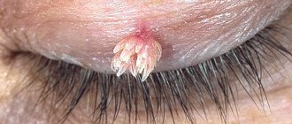
| Removal of 1 milia (grass, wen) in the eye area | 950 rub. |
| Removal of 1 wide-based skin lesion (digital dermatoscopy 750, removal 2150, anesthesia 550, histology 1500, free consultation on the day of treatment) | 4950 rub. |
| Removal of 2 wide-based skin lesions (digital dermatoscopy 750, removal 4300, anesthesia 1100, histology 3000, free consultation on the day of treatment) | 9900 rub. 9150 rub. ( 8% discount |
| Removal of 3 broad-based skin lesions (digital dermatoscopy 750, removal 6450, anesthesia 1650, histology 4500, free consultation on the day of treatment) | 14850 rub. 13350 rub. ( 11% discount |
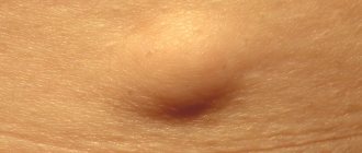
If you are reading these lines, then you probably have a formation (or several) on your skin and you want to get rid of them. keratoma pigment nevus fibroma milia (also called millet or wen)
hemangioma (angioma) dermatofibroma
- Do you think that keratomas or milia
on your face make you less attractive? - Because of a nevus
in your armpit, are you now wielding a razor like a professional hairdresser? - Are fibroids on your back
making it difficult to wear a bra? - Instead of playing sports and active recreation, are you thinking about accidentally damaging the hemangioma
? - At the beach, do you cover dermatofibromas
on your skin with a band-aid and are afraid to sunbathe? - Tired of touching the wart with your hand
or clothes, and are you thinking about getting rid of it for the hundredth time?
Read this text to the end and you will learn how to get rid of unwanted formations on the skin in one visit without scars and 100% safely.
Indications for surgical removal of a tumor
1. The indication for mole removal is the ABCDE test score:
- A – asymmetry of the neoplasm, uneven growth.
- B – fuzzy, blurry, jagged edges.
- C – heterogeneous color with blue, black, red splashes.
- D – large size, approximately a diameter of more than 5-6 mm.
- E – change in any of the characteristics (growth, pigmentation, surface relief).
If the mole begins to change in appearance, itching, swelling appears, and pain is felt on palpation, then you should immediately see a doctor.
2. Papilloma has a viral etiology and is the result of HPV infection. If the immune system cannot cope with the virus, then as a result of penetration and activation of a chronic infection, epithelium grows on the mucous membrane or skin. Not all papillomotosis is potentially dangerous, but there are strains of HPV that cause cell malignancy.
Oncogenic risk is carried by neoplasms that:
- They grow, increase in size, “spread”, their number increases.
- They darken, lighten, change color.
- They become denser and have a heterogeneous structure.
- Cause discomfort and pain.
It is recommended to remove any tumors that are easily injured due to their location. These can be papillomas (or moles) located on the neck, arms (especially on the elbow), and in women under the elastic bands of the bra. Large tumors on the lower back are also considered traumatic. When the skin is damaged, any infection easily penetrates through an open wound, creating more favorable conditions for inflammatory processes and suppuration. Therefore, it is recommended to remove potentially traumatic papillomas and nevi even if they do not bother you and do not carry an oncogenic risk.
3. Benign tumors have different etiopathogenesis. Some hormonally dependent forms (fibroids, etc.) require observation and are treated with medications (hormonal therapy), others are considered relatively harmless (fibroids), and others are considered precancer (for example, polyposis of the stomach and colon). Therefore, the treatment method is always chosen individually after a thorough examination and clarification of the diagnosis.
If benign tumors are detected, the nature of which cannot be determined, it is recommended to remove them surgically with histological examination of the excised tissue. Such neoplasms include tumors of the lymph nodes, internal organs and skin, which are externally similar to cystic formations or, for example, large inflamed lymph nodes.
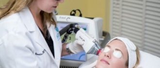
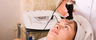
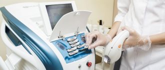
What is the essence of the radio wave surgery technique?
Recently, laser removal of skin tumors
sounds very often.
However, not everyone knows that there is a more modern method of removing skin lesions - radio wave surgery.
I will not bore you with the details of the physical foundations of the method. Let me just say that radiosurgery, just like laser
, is based on the evaporation of tissues that come into contact with the instrument.
In my opinion, these methods have more similarities than differences. However, compared to laser removal, radio wave exposure causes much less damage to surrounding tissue. This results in less inflammation in the area of removal,
shorter
healing times
and
excellent cosmetic results
.
Preparing for surgery
If a tumor is found in any part of the body, such as the breast, you need to know how to prepare for surgery.
Before removing a tumor, the patient must:
- get advice from an anesthesiologist;
- undergo a full examination of the body to identify possible contraindications to surgery;
- if you have chronic diseases, consult your doctor;
- take a biochemical blood test.
In addition, the patient may be prescribed antibiotics to prevent the development of an infectious process. You cannot eat on the day of surgery.
Benefits of removing skin lesions using radio waves
I am often asked “What is the best method to remove? Which is safer? Where is the best cosmetic result?
. Lately I have refrained from comparing methods because... the same best method may give different results in the hands of different doctors. That is why I will not talk about the advantages of the abstract radio wave method, but about the advantages of my original method of radio wave removal of skin formations:
- 100% safe removal.
Radio wave surgery allows you
to preserve the entire formation
for histological examination, regardless of its nature. The study is carried out in a certified laboratory by experienced morphologists. - Removal in 1 visit,
within a few minutes. Minimal damage to healthy tissue allows for the fastest possible healing. Re-inspection is not required or is optional. The wound is treated independently. - There are no restrictions
on sports and water procedures after removal. Suppuration in the area of operation is basically impossible.
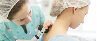
Progress of the procedure
For neoplasms such as fibroids, adenoma, abdominal surgery is performed. First, the doctor treats the skin with an antiseptic solution and makes an incision in the longitudinal or transverse direction. Having reached the organ, the surgeon examines it and removes the tumor tissue, being careful not to injure other organs. The resulting tumor mass is sent for histological examination.
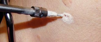
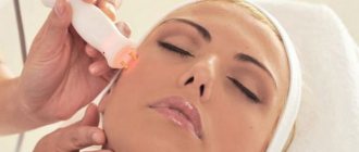
With the radio wave method of removal, the patient lies down in a chair, and the doctor treats the surgical site with an anesthetic. A special tungsten electrode is directed to the tumor. Under the influence of thermal energy, the epithelial tissues are heated, and the growth is removed to the root.
During cryodestruction, the patient lies down on a couch. The operation site is treated with brilliant green. A cotton swab or tip is cooled in nitrogen vapor and applied to the affected area.
Laser destruction is carried out under local anesthesia. The doctor punctures the tumor and dissects it with a laser knife. In this case, the laser beam touches the tissue and cauterizes the blood vessels, avoiding blood loss. The surgeon moves the dissected tissue apart, uses a special clamp to grab the tumor and separates it from the surrounding tissue.
Benefits not related to removal technique
- Honest prices.
No hidden fees or intrusive services. Either consultation or removal is paid, strictly according to the price list. You can always check the price with me on VKontakte by sending a photo with a ruler in the background. - Free information support before and after surgery.
If you have any doubts before your visit, you can easily and free of charge by mail or VKontakte. After the operation, you will have my personal phone number, by which you can ask me at any time about what is bothering you. - Fast and convenient.
Removal is carried out within a few minutes, in one visit. - Online appointment booking and removal without calls or waiting
- No preliminary tests, dressings, or suture removal required
- Histology results by email
. You can read them on these sites: docplanner.ru (more than 40 reviews), napopravku.ru (24 reviews), vk.com (more than 150 reviews)
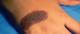
Photos before and after removal of skin lesions
Keratoma on the cheek
Fibroma on the face
Pigmented nevus above the lip
Nevus on the lower eyelid
26 more photos before and after deletion>>>
These advantages make radio wave surgery a leader in the removal of small skin lesions.
Summary:
As an oncologist, I can confidently recommend the radio wave surgery method for removing skin tumors. It allows you to examine all removed material and minimizes the risk of developing skin cancer and melanoma, and also leaves no traces for formations up to 5 mm in size.
Sign up for skin lesion removal OnLine right now! or by phone
Other articles:
- Removal of warts by a dermato-oncologist
- Removal of moles by a dermato-oncologist
- Safe mole removal? Radio knife!
- 16 symptoms of melanoma: what to do if you find one?
Useful article? Repost on your social network!
Treatment methods for benign tumors
The most reliable method of treating tumors is surgical removal of pathologically altered tissue. Since benign neoplasms do not grow into surrounding tissues and do not metastasize, removal is usually not particularly difficult. The exception is large tumors. For example, for some forms of uterine fibroids, hysterectomy is recommended - amputation of the organ along with the tumor.
To remove a benign tumor, various methods are used:
Surgical removal of tumor tumors
Radical removal of the tumor by excision within healthy tissue is the most reliable method of treatment. It allows you to completely remove a section of atypical tissue and stop tumor growth, eliminating the risk of malignancy (malignancy). Surgically removed tissue can be sent for histological examination to determine the type of tumor and clarify the diagnosis (benign or malignant).
With the surgical method of treatment, the tumor is excised - cut out, removing all altered tissues and cells. When small tumors are removed, the tissues are epithalized, completely or partially replacing the damage. When large tumors are removed, an additional second stage of treatment is later performed - cosmetic correction of the defect.
Surgical treatment can be used as the main method or in combination with antitumor therapy, immunotherapy, chemotherapy, hormonal therapy, etc.
With timely treatment, in most cases it is possible to achieve complete recovery and eliminate the risk of relapse.
The following types of benign tumors are removed surgically: fibromas, atheromas, wen, fibroids, etc.
Cryodestruction
Cryodestruction is another method of treating tumor tumors. Currently, liquid nitrogen is used for cryodestruction. Local freezing allows you to destroy the affected tissue areas, causing rejection of the tumor. Moreover, cryotherapy can be used both for superficial tissues and for deeper layers.
When exposed locally to the affected area, surrounding healthy tissue is not damaged. Cryonecrosis is the most gentle and virtually painless method of tumor destruction and does not require anesthesia. Exposure to cold blocks the vessels, making the removal bloodless, that is, bleeding does not occur.
The cryosurgical method of focal treatment with liquid nitrogen provides an anti-inflammatory and antiseptic effect, accelerates the regeneration process, and does not cause extensive defects and rough postoperative scars that require a subsequent cosmetic stage.
The method of cryodestruction removes papillomas, adenomas, genital warts, true erosions, warts, treats papillomotosis of the larynx, actinic keratosis, etc.
Electrocoagulation
Electrocoagulation is a method of removing atypical tissue areas using high-frequency electric currents. In a limited area, locally, with the help of an active electrode, a surgical field is created and the tumor tissue is destroyed using thermal exposure - subjected to destruction. The use of high-frequency currents eliminates the risk of infection and bleeding. However, burn damage to surrounding tissues is possible.
Using electrocoagulation, papillomas of viral etiology and moles are removed.