Dermatoscopy is an instrumental non-invasive study in which the doctor examines moles and other formations on the skin using a special device that creates multiple magnification. In oncology, dermatoscopy helps to distinguish harmless formations from skin cancer and identify melanoma in the early stages.
- Indications for dermatoscopy
- Why are moles dangerous?
- How is dermatoscopy performed?
- Equipment for dermatoscopy in the European Clinic
- Analysis of the received data
- Advantages of dermatoscopy using the FotoFinder system
- Prices for dermatoscopy at the European Clinic
Indications for dermatoscopy
- the appearance of any red, brown or black formation on the skin;
- injury to a mole; uneven change in color of the mole;
- an increase in the total number of moles and age spots;
- itching and tingling in the mole area;
- increase in size of the mole.
For preventive purposes, dermatoscopy is recommended:
- fair-skinned people with moles and age spots;
- with a family history of melanoma (if it was diagnosed in close relatives);
- people who have many moles and freckles on their body;
- when taking oral contraceptives for more than 1 year;
- when working in hazardous industries;
- with regular trips to hot countries;
- For dysplastic nevi, it is recommended to undergo dermatoscopy every six months to a year.
No special preparation is required for dermatoscopy. It is not recommended to use cosmetics or topical medications on the day of the examination. To obtain a clearer image, a small amount of gel is sometimes applied to the test site to reduce the reflection of light from the surface of the skin.
Modern digital dermatoscopes have many advantages over conventional ones. The device tube is connected to a computer monitor, on which the image is displayed. Digital dermatoscopy serves as a monitoring method for patients at high risk for developing melanoma. It also allows you to map moles over the entire surface of the body and observe their changes in dynamics, comparing the results with previous ones. The technique of digital dermatoscopy is simple, safe and fully automated. Within 3 minutes you can get a detailed analysis of all neoplasms.
It should be remembered that dermatoscopy does not allow a definitive diagnosis of melanoma to be made: this requires a biopsy and histological examination of a tissue sample.
What can you learn from the survey results?
Dermatoscope examinations are carried out by any private clinic. Therefore, if you suspect oncology, you can make an appointment with a dermatologist with a dermatoscopy service. There are no special contraindications to the study. Age restrictions and the presence of chronic pathologies do not affect the purpose of diagnosis.
The doctor is able to detect skin cancer at an early stage, when the compaction on the surface of the dermis does not cause suspicion among the patient and the therapist. If a dangerous disease is detected, the person is provided with medical assistance. This guarantees a positive prognosis for the patient with the exception of relapse.
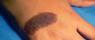
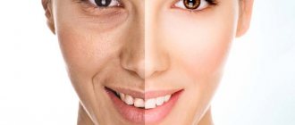
Photos of nevi through a dermatoscope
The service is considered affordable for all categories of the population. The price ranges from 300 rubles for the manual dermatoscopy method to 700 rubles. Computer - costs from 500 to 900 rubles. The epiluminescent method is considered the most expensive - the cost ranges from 700-1500 rubles.
A free service can be provided by a district clinic for a patient undergoing inpatient treatment or registered with a dermatologist. To conduct the study, you must register in advance and take the accompanying tests. The patient is sent to a state oncology center or skin clinic. Now there are many new dermatoscopes on the market - WelchAllyn (made in the USA), Heine from Germany and others.
Since the diagnosis is carried out visually, it does not require special preparation. The procedure is painless and does not require anesthesia. The main recommendation is to avoid applying creamy products to the examination site on the day of the doctor's visit. It is advisable to ensure the cleanliness of the skin being examined.
The procedure performed allows you to obtain more accurate information about the condition of the area being examined compared to simply a visual examination by a doctor. Based on the results of the study, the specialist receives information about the microstructure of the examined area and the formations existing on it.
Based on the data obtained after skin dermatoscopy, the doctor determines which of them may pose a danger. If any areas of skin with moles cause concern to the specialist, he may recommend surgical removal of the mole.
After the operation, the removed area is sent for histology for more detailed study.
The examination results are displayed by the computer program as a graph divided into several areas of different colors:
- a white tint means that the area being examined is in a safe area and does not pose a threat to the patient’s health;
- a yellow tint indicates that the mole does not need to be removed, but its condition must be regularly monitored;
- The red tint reflects the existing likelihood of the mole transforming into a malignant formation.
Based on the results of the data obtained, the specialist will offer several options. Having created a medical record, he will record the current condition of the patient and write down the results of dermatoscopy there. What this will require in the future and what additional measures the doctor will tell you immediately at the appointment.
The doctor may recommend surgery to remove the suspicious formation.
Why are moles dangerous?
Moles (pigmented nevi) are benign neoplasms, but some of them can transform into an aggressive, dangerous malignant tumor - melanoma. Risks are increased in the following conditions:
- A large number of moles on the body.
- Dysplastic nevi are special moles that are large in size, uneven edges, and uneven in color.
- Congenital melanocytic nevi are moles that are present on a child’s body from birth. Usually children are born without moles - they appear throughout life. The most dangerous are giant congenital nevi with a diameter of more than 10 cm. They degenerate into melanoma with a probability of 30%.
Important to know: Scientific research shows that only 30% of melanomas develop from pre-existing pigmented nevi. In 70% of cases, a malignant tumor occurs on unchanged skin, where there were no moles.
A Brief Review of the Best Dermatoscopes
Heine mini 3000 is a small pocket-type dermatoscope. Without replacing batteries, it can work for 10 hours. Lighting source - LEDs.
The peculiarity of the Heine Delta 20 hand-held device is that it can work both with and without immersion liquid (based on the principle of a polarizing dermatoscope). In addition, it is equipped with a contact board that allows connection to the camera. The device has lenses that provide 10x magnification.
KaWe Eurolight D30 is distinguished by rather large contact glasses (5 mm in diameter), the lenses provide 10x magnification. The lighting created by the halogen lamp can be adjusted. Another advantage of this device is a scale that allows you to determine the level of danger of pigmentation on the skin.
The Aramosg brand model is quite expensive, but also in demand on the market by dermatologists, cosmetologists and trichologists. In addition to traditional functions, the device can measure skin moisture levels, has special lenses for determining the depth of wrinkles and a built-in ultraviolet lamp for disinfection. This is a stationary type dermatoscope with the ability to connect to a computer or screen. The backlight in the device is adjusted automatically.
The Ri-derma device is more affordable in terms of cost than the previous model, but it is also more limited in functionality. This is a hand-held dermatoscope with 10x magnification lenses and halogen illumination. Can be powered by batteries or rechargeable battery.
Other popular dermatoscope options include DermLite Carbon and the miniature DermLite DL1, which can be connected to an iPhone.
Examination with a dermatoscope is a painless, quick, effective and inexpensive method to distinguish ordinary birthmarks and moles from malignant neoplasms. The main thing is not to delay a visit to a dermatologist if there is suspicious pigmentation on the skin.
More fresh and relevant information about health on our Telegram channel. Subscribe: https://t.me/foodandhealthru
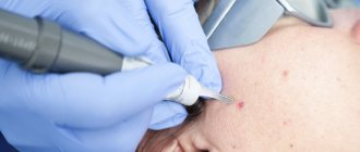
How is dermatoscopy performed?
Classic dermatoscopy is carried out using a special instrument resembling a magnifying glass - a dermatoscope. With its help, the doctor examines the skin and assesses the size and appearance of the detected tumors.
A more modern technique, which is used in the European clinic, is videodermatoscopy using the German PhotoFinder device. The device takes pictures of the entire surface of the patient's skin, creates a “mole map” and stores the images in a computer. The procedure is absolutely painless.
Devices - Why is FotoFinder a unique solution for dermatoscopy?
FotoFinder aesthetics
FotoFinder
Diagnostic complexes for diagnostics in aesthetic medicine, photo documentation, marketing of services
FotoFinder dermoscope Vexia
FotoFinder
Diagnostic complex for digital dermatoscopy combined with AI
FotoFinder ATBM bodystudio
FotoFinder
Diagnostic complex for body mapping, dermatoscopy and analysis
All Devices
Analysis of the received data
During dermatoscopy, a number of characteristics of formations found on the skin are assessed:
- Dimensions.
- Does the tumor rise above the skin?
- Shape, symmetry.
- State of boundaries: smooth/uneven, clear/blurred.
- Color, its uniformity.
- Character of the surface.
- The presence of ulcerations and other pathological changes.
In the PhotoFinder system, a computer program automatically analyzes images, thereby achieving greater diagnostic accuracy.
Subtypes of procedure
Depending on what type of dermatoscope is used during the manipulation, dermatoscopy is of several types:
- Digital (computer). Diagnosis of the formation is carried out using an electronic dermatoscope; research data is automatically displayed on the computer display. After evaluating the results obtained, the computer produces a printout with a conclusion in which the formation is assigned a certain level of danger on a scale from 0 to 100%. The advantages of such a study are the accuracy and speed of obtaining results, the absence of a subjective assessment of the doctor who performs the procedure.
- Manual. The examination of the skin is performed using a hand-held device that has the ability to magnify the image tenfold. The use of such equipment requires significant knowledge from the doctor, since a specialist must make a conclusion within 5-10 seconds.
This diagnostic manipulation allows you to determine the structure of pathological changes and conduct a differential diagnosis of any skin neoplasm. Based on the data obtained, based on the origin of the mole, the degree of its danger to human life, therapeutic tactics are determined.
Advantages of dermatoscopy using the FotoFinder system
- The diagnostic accuracy is 99%.
- While a dermatologist using a conventional dermatoscope does not examine the entire surface of the skin and may miss pathological neoplasms in hard-to-reach places, the FotoFinder system provides the most complete scanning of the body surface.
- The ability to detect melanoma and other small skin tumors in the early stages.
- If videodermatoscopy is performed regularly, the doctor is able to assess the condition of the skin over time, and this further increases the accuracy of diagnosis.
- The system can automatically analyze images.
- The study is completely safe, painless, and has virtually no contraindications.
Dermatoscopy of skin tumors. Dermatoscopy of skin and moles
Every person has moles. Also, other neoplasms often arise that begin to bother you. The question arises of where to check the nature and danger of a cutaneous nevus. Moles can also increase in size. A dermatologist studies the clinical nature of nevi. Dermatoscopy is a scientific method of studying skin formations for internal structure and degree of malignancy. The method allows for early detection of cancer.
Characteristics of the method
Dermatoscopy refers to instrumental methods for examining the skin of the human body. For this purpose, a special device is used - a dermatoscope. This method is used by many doctors - cosmetologist, dermatologist and oncologist.
Dermatoscopy is a modern diagnostic method. Before the development of the technique, histology was used to examine suspicious formations on the upper layer of the dermis. Partial excision of the nodule was carried out with further examination for the presence of malignant cells. This technique is quite painful for humans and is accompanied by certain risks of complications - it can provoke rapid growth and infection of the nevus.
A dermatoscope allows you to enlarge a suspicious area (enlarged nevus or inflammation) and study the structure of the neoplasm in detail. The non-invasive method is distinguished by accuracy and accessibility. Error in the study is excluded.
Diagnostics helps to identify:
- Enlarged nevus, papillomas with warts;
- Change in color of the neoplasm associated with cell degeneration;
- The structure changes density;
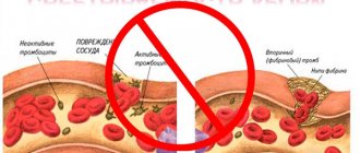
- Capillaries located nearby are injured, microbleeding occurs;
- The skin around the mole and other formations changes color and cracks appear.
The main principle of using the procedure is to detect skin cancer (melanoma) in the early stages. Dermatoscopy of moles helps to detect the presence of pathologies on the skin of melanocytic and non-melanocytic formations. Pigmentless melanoma, detected at an early stage of development, is easy to treat and guarantees recovery in 90% of patients.
The procedure is often prescribed for:
- Determining the results of treatment;
- Determining the correct method of excision of a dangerous formation;
- Establishing a course of treatment for the degeneration of a mole;
- Differentiation of diseases of the scalp;
- Diagnosis of nail plate pathology;
- Controlling the development of nevi.
The formation of nodules suspected of being malignant is studied by an oncodermatologist using a dermatoscope.
Types of dermatoscopy
Dermatoscopic examination can be carried out in various ways. Currently there are:
- The manual method of dermatoscopy allows you to study the structure of the neoplasm due to tenfold magnification at the maximum depth into the layers of the dermis. A simple and informative method is often used in clinical practice. It is carried out with and without a camera. With a camera, the pictures are displayed on the screen and then printed for attachment to the medical record. It is convenient to analyze the trend of nevus development using the images. If a camera is not available, the doctor will make the test results.
- Digital dermatoscopy of skin tumors makes it possible to determine the size, structure and boundaries of the suspicious area. Medical examination is carried out using a computer with
- Epiluminescent dermatoscopy is the latest technique in medicine. Based on the use of polarized lighting. Shows the tumor from the inside. The accuracy of diagnosis is 95%.