There are two conditions that fall under the concept of "malignant mole" . The first is melanoma- dangerous neoplasms , which are initially prone to malignancy (congenital melanocytic nevus and dysplastic nevus). The second is the immediate beginning of malignancy of the mole , when cancer cells have not yet grown into the underlying tissues and have not metastasized. The second condition is also called “cancer in situ,” which means a local malignant tumor that has not spread throughout the body.
Photo 1. A mole is a congenital safe neoplasm that, under unfavorable conditions, can degenerate into cancer. Source: Flickr (maria—-).
Are colorless moles on the body dangerous?
Any growths on the body are a pathology of the epidermis, and a natural phenomenon. Benign neoplasms do not cause trouble or problems to their owner. But under the influence of certain factors, it degenerates into a malignant form.
The nature of colorless moles that do not pose a threat to health can be determined by certain signs:
- the edges are smooth and clear;
- pliable, not rigid;
- absence of inflammatory processes around the growth;
- small size;
- do not protrude much above the surface.
In other cases, flesh-colored neoplasms indicate the presence of serious diseases, such as Vitiligo, Leukoplakia, Seborrhea.
A transparent mole occurs on the body in single and multiple cases. Colorless moles are localized mainly on the face, forehead, neck, head, chest, and armpits.
Several types of colorless neoplasms are noted.
| Form | View |
| Flat | A birthmark with a smooth surface. It looks like a skin spot that gradually discolors. |
| Warty | Neoplasms are formed from birth. Their size can reach 20 mm. They often focus on the nose and eyelid. |
| Hanging | Convex small growths can most often be observed on the neck. They do not cause inconvenience, they look like berries. |
Some colorless birthmarks are located in dangerous areas where they can easily be touched or injured. You need to handle them carefully so as not to cause degeneration into oncology.
Possible complications
If the formation does not change and does not show any signs, then complications do not threaten. A nevus becomes dangerous when its asymmetry is observed, it acquires a heterogeneous color, and its size increases (more than 6 mm). A pink mole can be dangerous if it is damaged. There is a high probability of its degeneration into melanoma (malignant tumor), which has the following features:
| Risks of occurrence | Features of manifestation | Treatment and prevention |
|
|
|
Reasons for appearance
Colorless moles do not appear spontaneously. One can always find a logical source for their origin.
Medical practice identifies several etiotropic factors that influence the formation of colorless growths:
- long-term exposure to aggressive ultraviolet rays;
- hormonal surges associated with pregnancy and puberty;
- chronic inflammatory disease of the sebaceous glands;
- invasive cosmetics;
- lack of vitamins and minerals;
- taking photosensitizing drugs;
- a symptom of calcium deficiency and vitamins B and E;
- mechanical damage to the epidermis;
- heredity;
- taking hormonal drugs.
Sometimes even bad habits can lead to the appearance of colorless growths. Therefore, you should reconsider your lifestyle.
Often, ordinary new growths can fade as the process of aging and death occurs. But if the area around them becomes lighter, you should urgently consult a doctor.
An important point is to independently or with the help of specialists determine the cause of the appearance of colorless neoplasms in order to prevent the development of others. This is especially true for women, since even a colorless mole on the body or face leads to a cosmetic defect.
Diagnostics
In order to determine the specific type of nevus, the following diagnostic measures are used:
- Dermatoscopy. It is carried out to study the depth of the tumor, its contours and structure.
- Visual inspection of the mole.
- Histological examination allows one to accurately determine the type of nevus. It is carried out only after the excision process is completed.
- Ultrasound. It is necessary to study the infiltrative growth of the formation and its depth.
- Siascopic examination. With its help, doctors determine the degree of distribution of pigment in the cells, which forms the formation itself.
The traditional way to diagnose a nevus is its excision and subsequent examination, but thanks to dermatoscopy it is possible to determine the type of mole without intervention
Note. Blue nevus is visually noticeably different from other types, but a full diagnosis is still necessary to determine the depth and structure of the neoplasm.
If we consider the clinical and morphological characteristics of blue nevus, we can distinguish three key types:
- Cellular . The size of such a mole can vary from 1 to 3 cm. The surface itself is rough, and the shape resembles an hourglass. The color is most often dark blue. The external signs of such a nevus resemble melanoma. Most often, this type is recorded in the buttocks or lower back. Much less often it forms on the back of the feet and hands.
- Combined . This form of blue nevus of the skin is distinguished by the fact that it combines the characteristics of a standard and borderline mole, and sometimes even a complex segmental formation.
- Simple . In this case, the size of the mole is limited to 1 cm, and its surface is smooth. The most common shape is round. The color can vary from dark blue to light gray. Moles of this form are found mainly in the neck, on the arms, face, and less often on the oral mucosa.
Cellular nevus is the largest, which frightens people and forces them to see a doctor
Note. Blue nevus can be either single or appear in many places on the body.
Signs of colorless moles that need to be seen by a doctor
A benign flesh-colored mole does not often change into a threatening shape. Oncology can begin due to excessive production of melanocyte cells. The most common negative consequence is melanoma.
Serious signs of colorless neoplasms that require special attention include:
- the appearance of a white halo;
- if a small birthmark grows;
- presence of plaque on the surface;
- there was pain when touched;
- began to bleed;
- the presence of white inclusions;
- the mole has become discolored;
- It started to get dark.
If the corresponding symptoms are present, you should immediately consult a doctor. A specialist will carefully examine and find out how the colorless growth began to appear. A dermoscopic analysis is required, which shows whether the pigmented formation needs to be eliminated or not.
You should contact a dermatologist or surgeon if you develop new colorless birthmarks, as well as when modifying existing ones. Modern equipment allows you to track the dynamics of the development of a mole and measure the level of pigment. The specialist will determine the nature of the tumor and, if necessary, prescribe therapy.
A person who is at risk needs to be careful on the beach and avoid mechanical injuries.
Types of blue nevus
Medical practice divides blue nevus into three subtypes:
- Simple - no more than 1 cm, with a smooth surface and dense in consistency. It can appear on the body (usually the hands), face (including the décolleté area) and mucous membranes (uterus, vagina, oral cavity).
- Cellular - can reach sizes up to 3 cm, has a dark blue color with irregularities. For this subspecies, the typical location is only on the body (lumbar region, buttocks, hands and feet).
- Combined - in this case, the blue nevus is combined with other types of dangerous and non-dangerous moles.
Features of removing colorless moles
Almost a third of skin cancer arises from degenerated growths. The diagnosis can only be made by a doctor. However, you need to pay more attention to the color change. And if the tumor turns black, this signals that the tumor cells begin to divide chaotically and produce the pigment melanin. But there are colorless melanomas, which even for a specialist are difficult to distinguish from benign skin pathologies.
Many people are afraid to get rid of colorless birthmarks, thinking that they will harm themselves. There are a number of moles, so-called special multiple dysplastic growths. They must be removed for preventive purposes. They have a very high risk of degenerating into a malignant form.
Colorless growths should be removed only in specialized institutions. Having taken genetic material for research, the doctor determines whether the operation can be performed and what is the best way to do it.
Modern medicine offers different options for implementing this process. Colorless neoplasms are excised, as are pigmented birthmarks.
| Type of operation | Description |
| Surgical method | The manipulation is performed under local anesthesia. This is a popular method that is used to excise large growths. However, this method can remove all moles, regardless of the depth of germination. At the end of the operation, cosmetic stitches are applied. After a week they are removed. Light scars remain. |
| Electrocoagulation | The session begins with disinfection of the birthmark area. The operation is performed with special equipment using an electrocoagulator under local anesthesia. The tissue is being cauterized by electric current. The electrode heats up and burns out the tumor. After the operation, a crust remains, which protects the wound from infection and bleeding. |
| Chemical destruction | The destructive effect is caused by chemicals such as solcoderm, cantharidin, salicylic, and lactic acid. The substance is applied to the growth. After a few days, a dry crust forms, which then falls off. The wound heals in 30 days. |
| Cryodestruction | Freezing pathological tissues with liquid nitrogen. Under the influence of low temperature, the blood supply to the birthmark is disrupted, which leads to the destruction of the affected tissue. This operation occurs quickly and without pain. |
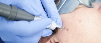
| Laser method | The operation involves directing a laser beam at the tumor. A layer-by-layer removal of the skin area on the birthmark occurs. After 14 days, complete healing of the wound occurs, leaving no trace. |
| Radio wave surgery | A non-contact method where high-frequency waves of thermal energy cut tissues and remove pigmented cells. |
All methods do not require hospitalization. But after the operation, preventive measures should be taken.
Is it possible to bleach a mole yourself?
Many women are concerned about the question of how to discolor a mole on their face. Some try to whiten using traditional cosmetology recipes. Several methods are used to lighten birthmarks. All of them are based on the application of lotions soaked in certain products, such as:
- lemon juice;
- tomato juice;
- olive oil;
- vitamin A;
- vitamin E;
- Apple vinegar;
- iodine;
- oats with milk and rice flour.
To make moles lighter, it is recommended to spend less time in the sun and use sunscreen.
Any independent manipulation of birthmarks is very risky. It is necessary to monitor moles and, if they transform, urgently contact a dermatologist.
Treatment methods
Surgical removal
Surgical removal involves the following basic methods:
- Cauterization, which is used extremely rarely because it is not particularly effective. This is explained by the fact that most of the nevus is located in deep layers, and the cauterization method affects only the top layer. With such removal, there is a high probability of a new mole appearing.
- Laser removal is an effective method for pink mole removal.
- Infrared or light coagulation is also used.
It is easier and faster to remove flat moles. When removing a dangerous or unwanted formation, various anesthetic creams are used, since the procedure is not particularly pleasant. Depending on the size and type of lesion, removal may leave a small red spot that will go away over time. In the first month after removal, it is not recommended to visit a solarium or be in the open sun.
Traditional therapy
If the formation is small in size and benign, then you can try to remove it using folk remedies. Since such therapy can be harmful, you should consult a specialist before using it. Common folk methods for getting rid of pink moles:
- Using vinegar to smear the nevus before bed.
- Mix garlic, vinegar and honey, apply the resulting mixture to the mole and leave for 4 hours. The treatment lasts a month, and the procedure is carried out every day.
- Celandine juice, which is applied to the damaged area of the skin 2 times a day, is considered effective.
- An ointment consisting of dandelion roots and butter. The medicine is applied to the pink mole three times a day.
- To remove a raised nevus, use pineapple juice.


