How to hide moles on your face? In all centuries, fashion has dictated its own laws. If 100 years ago a spot on the face was a sign of some kind of coquetry, then modern women are seriously solving the problem of removing formations on the face.
Of course, if the nevus is in the “right” place, it still looks very good. But there are also “wrong” places where it is advisable to remove them. “Right” and “wrong” places are regulated by fashion. Any woman can name famous models who are famous precisely because of the nevi on their faces.
However, not every mole can be removed surgically. And the point is not that this is technically impossible, everything is very good with this in the modern world. The fact is that some nevi are too close to nerve nodes or blood vessels and it is strictly not recommended to touch them.
But there is no need to despair, modern skin care products and means of concealing imperfections on it are developing by leaps and bounds. To hide a mole on your face, you need to perform several simple manipulations with cosmetics.
Are colorless moles on the body dangerous?
Any growths on the body are a pathology of the epidermis, and a natural phenomenon. Benign neoplasms do not cause trouble or problems to their owner. But under the influence of certain factors, it degenerates into a malignant form.
The nature of colorless moles that do not pose a threat to health can be determined by certain signs:
- the edges are smooth and clear;
- pliable, not rigid;
- absence of inflammatory processes around the growth;
- small size;
- do not protrude much above the surface.
In other cases, flesh-colored neoplasms indicate the presence of serious diseases, such as Vitiligo, Leukoplakia, Seborrhea.
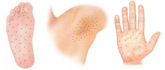
A transparent mole occurs on the body in single and multiple cases. Colorless moles are localized mainly on the face, forehead, neck, head, chest, and armpits.
Several types of colorless neoplasms are noted.
| Form | View |
| Flat | A birthmark with a smooth surface. It looks like a skin spot that gradually discolors. |
| Warty | Neoplasms are formed from birth. Their size can reach 20 mm. They often focus on the nose and eyelid. |
| Hanging | Convex small growths can most often be observed on the neck. They do not cause inconvenience, they look like berries. |
Some colorless birthmarks are located in dangerous areas where they can easily be touched or injured. You need to handle them carefully so as not to cause degeneration into oncology.
What moles can be removed
Owners of such moles are extremely attractive to the opposite sex.
In ancient times, it was believed that a person with a mole on the lip (both a man and a girl) was kissed by the goddess of love herself and good luck would accompany him or her. Such people have many friends, they easily form relationships and are rarely lonely. Men adore girls with moles on their mouths (remember Marilyn Monroe!). At the same time, a mole near the lips is most often a sign of a monogamous person in both men and women (unless it is in the corner of the mouth, because the meaning of such a mark is a tendency to flirt). There are also many workaholics among such people; perhaps, love for work and love for a person are almost the same thing for them.
https://www.youtube.com/watch?v=4q_FgHF7-II
Those who have dark spots on their skin should be wary of their changes. In time, detected signs of degeneration into melanoma contribute to the timely removal of the formation and preservation of health. Safe moles are different:
- the presence of a stalk – it cannot be formed by malignant cells that grow randomly;
- long-term condition without changes.
Spots that appear shortly after birth are not considered dangerous. It is important that they are small in size. Good – non-dangerous – signs of neoplasms include:
- flesh tone;
- unchanged pattern of the skin of the nevus and adjacent tissues;
- soft consistency;
- hair on the surface of the neoplasm - growing from the skin, indicates the absence of pathologies;
- diameter no more than 5 mm;
- symmetry;
- nevus in the form of a spot.
Why do people with nevi on their bodies need to monitor their changes? There is always a threat of degeneration of non-dangerous tumors into a cancerous tumor. What moles are dangerous to health? Key signs you need to know:
- change in shades towards the dark side, the appearance of multi-color;
- rapid increase in size - exceeds two millimeters per year;
- occurrence of cracks;
- the formation of asymmetry due to uneven growth;
- lack of elasticity;
- the appearance of itching, burning;
- presence of discomfort.
The appearance of dangerous moles requires an immediate visit to a specialist to clarify the nature of the changes and the likelihood of developing skin cancer. Pathological transformations provoke:
- injury to the nevus due to negligence;
- self-removal;
- abuse of exposure to the sun, use of a solarium;
- location of the formation in places of frequent contact with clothing - on the neck, head, genitals, legs;
- placement in the hair, on the face, palms - where there is a high probability of injury;
- previously removed melanoma.
Not a single person is protected from the sudden proliferation of cells of a harmless mole. Melanoma is an extremely serious disease. Changes not detected at the initial stage can result in death. The provoking factor is unsuccessful independent removal of tumors. Moles are dangerous because of their ability to:
- transform into an atypical – precancerous form;
- grow to large sizes;
- turn into cancerous;
- with minor external changes, metastases actively spread throughout the body through the circulatory and lymphatic channels.
What do nevi that are subject to pathological changes look like? Only a doctor can correctly distinguish between non-dangerous tumors. Dangerous formations look like this:
- blue – seals under the skin with clear boundaries, with dimensions no more than 10 mm;
- nodal – round, flat in shape, color – brown, black;
- skin – often pale, convex;
- halo nevus - pigment surrounded by a light or white rim;
- Spitz - looks like a dome-shaped tumor of pink shades, with the possible presence of a hole through which blood and fluid leaks;
- connective - connect individual formations into a whole.
One of the signs of a non-hazardous formation turning into a dangerous one is a change in contours. It often has blurred edges and scalloped borders. There are non-dangerous types of nevi - dysplastic. Only a specialist can make a correct diagnosis. A mole with uneven edges can be dangerous if there are additional signs of melanoma:
- accelerated changes in size;
- the presence of clearly defined asymmetry;
- the appearance of highly indented boundaries.
Rough mole
Such a neoplasm is harmless if its diameter is no more than 5 mm and remains constant in size. Often its appearance signals a lack of vitamins and nutritional disorders. Doctors advise coming for a consultation if it is discovered that:
- the smooth nevus turned into a rough one;
- bothered by burning, itching, tingling;
- irregularities and compactions appeared in the middle;
- areas with different shades formed;
- diameter has increased significantly.
A dangerous rough mole requires immediate examination if:
- the appearance of bleeding;
- development of the inflammatory process;
- rapid change in size;
- formation of asymmetry;
- formation of purulent discharge;
- the occurrence of painful sensations when touched;
- the emergence of an irregular shape, blurred boundaries, along the edges of the neoplasm.
Large moles
Large formations on the skin are pigment spots. When they remain unchanged and do not cause inconvenience, this is not a dangerous phenomenon. It is important to constantly monitor their appearance, color, and size. To eliminate worries, you need to consult a dermatologist. During the visit, the specialist will conduct a diagnosis and give a forecast of the risk of developing a malignant neoplasm. Large moles become dangerous if they:
- injured;
- thickened;
- started to itch;
- were unsuccessfully removed independently;
- changed in size, shape;
- are bleeding.
Often nevi cause trouble for women when they are in a visible place - the face, neck. Even if they do not bother you, using removal will be the right decision - the appearance will improve significantly. After the procedure, the doctor must send the tissue for histological analysis to decide whether the mole is malignant or not. If the neoplasm is not dangerous, does not bother you, and does not change in size, surgery is not required. What moles cannot be removed? Experts believe:
- there are no contraindications;
- It is important to choose the right excision technique.
You should be careful about skin growths; it is unacceptable to remove them yourself. Only the doctor will determine whether a nevus is dangerous or not and decide what to do with it. You can delete it if:
- injured from clothing - on the neck, in the groin area, under the armpits;
- cause pain when touched;
- are located under the hair on the head and can be damaged when combing or cutting;
- change color, shape, outline;
- significantly increase in size;
- characterized by the presence of burning, itching;
- accompanied by inflammation and bleeding.
Nevi are essentially benign skin formations.
Some people get scared when they see many moles on their body. What does this mean for a specialist? Only that the patient’s body is prone to accumulation (accumulation) of melanin in the surface layers of the epidermis.
1. External:
- exposure to ultraviolet radiation, one of the most common factors in the formation of moles due to increased melanin levels during tanning;
- traumatic damage to the epidermis, systematic violations of the integrity of the skin contribute to the appearance of pathological changes in it;
- exposure to radiation, which rapidly changes normal skin cells;
- consumption of harmful products (GMOs, fast food, alcohol) and smoking, these habits negatively affect metabolic processes in the body.
2. Internal:
- endocrine disorders and diseases, any changes in hormonal levels can cause the appearance of skin pathologies, pigmentation, moles;
- hereditary predisposition, the presence of various nevi in the family.
The appearance of moles can be triggered by inflammatory and autoimmune skin diseases, toxic lesions, burns and frostbite, as well as the constant use of low-quality cosmetics and preparations for face and body care.
Ordinary nevi do not cause any discomfort to their carriers; sometimes they even disappear without a trace. Their shape is stable, size and color remain unchanged.
But benign moles are sometimes removed to prevent their degeneration if they are of rather large size (pedunculated) and are located in areas of the body where a person constantly injures them (during vigorous activity or parts of clothing).
Remember dangerous moles in the photo and be vigilant!
If nevi degenerate into cancerous moles, this is dangerous, since such neoplasms quickly metastasize to other organs.
1. The shape of the mole changes, it loses its symmetry and begins to grow in one direction.2. The edges of the nevus become uneven (“ragged”, “torn”).3. The color of the mole is uneven and contains yellow, red or black inclusions.4. The nevus grows or “shrinks”, its size changes rapidly.5.
The texture of the mole becomes different, smooth turns into rough, bumpy into flat, etc.6. Loss of hair growing from the nevus.7. Itching, peeling and burning in the area of the mole. There are several reasons why a nevus itches: – pathological cells multiply; – active processes of healthy tissue dying are underway; – the area around the formation becomes inflamed and swells.8. The appearance of microcracks and ulcerations.9. Bleeding and soreness of the mole.
Cancerous moles (melanomas): photo
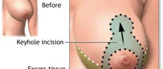
Many people want to get rid of birthmarks and nevi not only in cases of malignancy (pathological change), but also to correct a cosmetic defect.
However, removal is carried out at the oncology center for people with malignant tumors and with a high probability of degeneration of nevi.
Often the indication for surgery is the localization of moles on the body: on the scalp, on the neck, on the chin, in the area of the shoulder blades.
Methods for removing nevi (with information on how much it costs to remove them):
- surgical, using local anesthesia and a scalpel (in municipal surgical hospitals, according to indications - free of charge, in medical centers from 300-500 rubles)
- cryofreezing with liquid nitrogen (1000-1500 rubles);
- electrocoagulation - cauterization with electric current (600-1300 rubles);
- photodynamic - ultraviolet irradiation (1000-1200 rubles);
- laser - removal of moles with a beam (800-2000 rubles);
- radio wave, destruction of nevi by shock radio wave (RUB 700-1400)
- Do not use products containing alcohol.
- Do not try to hide reddened skin with makeup.
- Refrain from going to the solarium and do not sit in the sun.
- Don't tear off the crust; it should fall off on its own.
Reasons for appearance
Colorless moles do not appear spontaneously. One can always find a logical source for their origin.
Medical practice identifies several etiotropic factors that influence the formation of colorless growths:
- long-term exposure to aggressive ultraviolet rays;
- hormonal surges associated with pregnancy and puberty;
- chronic inflammatory disease of the sebaceous glands;
- invasive cosmetics;
- lack of vitamins and minerals;
- taking photosensitizing drugs;
- a symptom of calcium deficiency and vitamins B and E;
- mechanical damage to the epidermis;
- heredity;
- taking hormonal drugs.
Sometimes even bad habits can lead to the appearance of colorless growths. Therefore, you should reconsider your lifestyle.
Often, ordinary new growths can fade as the process of aging and death occurs. But if the area around them becomes lighter, you should urgently consult a doctor.
An important point is to independently or with the help of specialists determine the cause of the appearance of colorless neoplasms in order to prevent the development of others. This is especially true for women, since even a colorless mole on the body or face leads to a cosmetic defect.
Dysplastic nevus is a strange mole.
A dysplastic nevus is any mole that deviates from the standard appearance for such formations. For example, it has blurry edges or an irregular shape. Heterogeneous color from light brown to red-brown. Including the diameter of the mole, reaching 1.4 cm. On the one hand, the listed changes may be a sign of melanoma. On the other hand, these signs are not so pronounced. It is not always possible to distinguish a dysplastic nevus from cutaneous melanoma. Therefore, it is advisable to remove all such moles and observe them, at a minimum. The formation of dysplastic nevi usually occurs during or before puberty. Cases of occurrence throughout life cannot be excluded. Changes in moles are often associated with excessive sun exposure. Frequent locations of this mole: torso, buttocks, limbs, scalp, face, genitals. But it may also appear on other areas of the skin. The number of such moles varies up to 100. In the center of the nevus there is sometimes a darker area. In addition, such a mole may not only be similar to skin melanoma, but over time it may degenerate into one, especially if there is a family predisposition.
Borderline nevus of the skin of the palm. Do not injure this mole. Keep away from the sun and observe.
Another flat mole. However, the center is darker in color than the periphery. This allows us to attribute it to dysplastic nevi.
Signs of colorless moles that need to be seen by a doctor
A benign flesh-colored mole does not often change into a threatening shape. Oncology can begin due to excessive production of melanocyte cells. The most common negative consequence is melanoma.
Serious signs of colorless neoplasms that require special attention include:
- the appearance of a white halo;
- if a small birthmark grows;
- presence of plaque on the surface;
- there was pain when touched;
- began to bleed;
- the presence of white inclusions;
- the mole has become discolored;
- It started to get dark.
If the corresponding symptoms are present, you should immediately consult a doctor. A specialist will carefully examine and find out how the colorless growth began to appear. A dermoscopic analysis is required, which shows whether the pigmented formation needs to be eliminated or not.
You should contact a dermatologist or surgeon if you develop new colorless birthmarks, as well as when modifying existing ones. Modern equipment allows you to track the dynamics of the development of a mole and measure the level of pigment. The specialist will determine the nature of the tumor and, if necessary, prescribe therapy.
A person who is at risk needs to be careful on the beach and avoid mechanical injuries.
A large cauliflower-shaped mole is a papillomatous nevus.
Papillomatous nevus is one of the most common moles. It has an irregular shape, a bumpy surface that rises above the skin level. Color from flesh to brown. It rarely happens that a mole is black in color. It is located mostly on the face, neck, and scalp, but can be located on any part of the body. There is a gradual growth of the mole, which reaches large sizes. Being on the head, face, neck, it is often injured, as a result of which it becomes inflamed and painful. Pigmented hair grows on it and makes the mole a significant and major cosmetic defect. Frequent injury, scratching of the skin, and psychological factors serve as reasons for removing the tumor. This happens using a laser, liquid nitrogen, or radio wave method. After removal of a mole, histological examination is sometimes required to rule out melanoma.
Features of removing colorless moles
Almost a third of skin cancer arises from degenerated growths. The diagnosis can only be made by a doctor. However, you need to pay more attention to the color change. And if the tumor turns black, this signals that the tumor cells begin to divide chaotically and produce the pigment melanin. But there are colorless melanomas, which even for a specialist are difficult to distinguish from benign skin pathologies.
Many people are afraid to get rid of colorless birthmarks, thinking that they will harm themselves. There are a number of moles, so-called special multiple dysplastic growths. They must be removed for preventive purposes. They have a very high risk of degenerating into a malignant form.
Colorless growths should be removed only in specialized institutions. Having taken genetic material for research, the doctor determines whether the operation can be performed and what is the best way to do it.
Modern medicine offers different options for implementing this process. Colorless neoplasms are excised, as are pigmented birthmarks.
| Type of operation | Description |
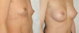
| Surgical method | The manipulation is performed under local anesthesia. This is a popular method that is used to excise large growths. However, this method can remove all moles, regardless of the depth of germination. At the end of the operation, cosmetic stitches are applied. After a week they are removed. Light scars remain. |
| Electrocoagulation | The session begins with disinfection of the birthmark area. The operation is performed with special equipment using an electrocoagulator under local anesthesia. The tissue is being cauterized by electric current. The electrode heats up and burns out the tumor. After the operation, a crust remains, which protects the wound from infection and bleeding. |
| Chemical destruction | The destructive effect is caused by chemicals such as solcoderm, cantharidin, salicylic, and lactic acid. The substance is applied to the growth. After a few days, a dry crust forms, which then falls off. The wound heals in 30 days. |
| Cryodestruction | Freezing pathological tissues with liquid nitrogen. Under the influence of low temperature, the blood supply to the birthmark is disrupted, which leads to the destruction of the affected tissue. This operation occurs quickly and without pain. |
| Laser method | The operation involves directing a laser beam at the tumor. A layer-by-layer removal of the skin area on the birthmark occurs. After 14 days, complete healing of the wound occurs, leaving no trace. |
| Radio wave surgery | A non-contact method where high-frequency waves of thermal energy cut tissues and remove pigmented cells. |
All methods do not require hospitalization. But after the operation, preventive measures should be taken.
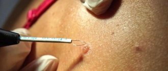
What happens if you rip off a mole?
You cannot remove a nevus yourself.
Firstly, it is dangerous, and secondly, it is simply ineffective. If the mole is located close to blood vessels, prolonged bleeding may develop. Often such self-medication leads to re-formation of the nevus, its growth or malignancy.
Therefore, it is difficult to predict what will happen if a mole is torn off. It may have no consequences, or it may cause serious complications for the health and life of patients.
Doctors warn that any trauma to the nevus is extremely undesirable, but pedunculated moles or small convex formations can be accidentally removed with nails or hard items of clothing.
What to do if you rip off a mole:
- cauterize the wound with an alcohol solution;
- stop the bleeding by applying a gauze bandage;
- come to see a specialist.
In cases of partial removal of a mole, do not touch the remaining formation, do not cut it off or tear it off.
If a white spot , only a dermatologist can determine this. However, if you have a mole with a white halo, this may mean that the neoplasm has begun to degenerate into melanoma, which must be prevented as soon as possible.
Sometimes such pigmentation appears before the disappearance of a mole (as a sign of depigmentation), and in other cases it may signal its degeneration. White spots around the mole appear as a characteristic sign of Setton's nevus. This formation is considered harmless in terms of degeneration into a more malignant form, however, melonomas (aggressive cancerous formations) can also have such a white rim, so the appearance of white spots is a reason to contact a medical specialist.
Is it possible to bleach a mole yourself?
Many women are concerned about the question of how to discolor a mole on their face. Some try to whiten using traditional cosmetology recipes. Several methods are used to lighten birthmarks. All of them are based on the application of lotions soaked in certain products, such as:
- lemon juice;
- tomato juice;
- olive oil;
- vitamin A;
- vitamin E;
- Apple vinegar;
- iodine;
- oats with milk and rice flour.
To make moles lighter, it is recommended to spend less time in the sun and use sunscreen.
Any independent manipulation of birthmarks is very risky. It is necessary to monitor moles and, if they transform, urgently contact a dermatologist.
Concealing with foundation
The most effective way to disguise nevi on the face is foundation. Its texture is quite heavy, but it is easy to work with. And the feeling of using it is quite pleasant. At the very least, the cream is definitely not capable of causing discomfort. Of course, if you don't overdo it.
Since the cream is quite thick, to hide a mole, you need to apply 1 layer. There is no need to strive for the best by applying several more layers. The fact is that no matter how good the cream is, it will not be possible to achieve a complete match of skin tones and foundation, and its abundance will be striking. In this case, you will have to cover the entire surface of your facial skin with foundation, and this is completely unnecessary. The cream is applied with a special brush directly to the mole itself. In this case, the product should not cover the skin around the mole.
Brown papillomas are like moles.
Pigmented papillomas are benign skin tumors caused by a virus. The change in color to light or dark brown makes them very similar to nevi and seborrheic keratoses. Accordingly, they are also often called “moles.” They look like a papilla on a thin stalk. It tends to spread to other areas of the skin over time. Infection occurs through contact and household contact. The impetus for its appearance is a weakened immune system, bad habits, and pregnancy. Treatment of moles involves removing papillomas with a laser, cryodestruction, or radio wave method. Which is best combined with taking antiviral and immunostimulating drugs.
Rough moles are filamentous warts.
Warts on the body actually come in several varieties. Most people consider warts only those that are located on the hands of teenagers - these are ordinary warts. However, there are also filiform, plantar, and periungual. And in the groin and anus - genital warts. Patients can refer to moles as thread-like warts that appear on the torso, shoulders, forearms, thighs and legs. Despite their name, they are not at all thin and long, and do not in any way resemble a thread. Simply, they often have several finger-like horny outgrowths on their surface. However, modern cosmetics (even soap) help to quickly soften the horny masses and remove (wash off) the outgrowths. All that remains of a filamentous wart is a rough, protruding base, which can easily be called a mole. The appearance of such moles is promoted by the human papillomavirus. It is transmitted from another person, being contagious. Size from 1-3 cm in diameter, uneven surface, dry and rough to the touch, often painful. The impetus for the appearance of moles is stress, wearing clothes made of synthetic fibers, wearing shoes that are too tight, hypothermia, and excessive sweating. Removal is recommended to prevent the spread of human papillomavirus, which contributes to cancer. Sometimes, these moles can disappear on their own within a couple of months or years.
Brown rough moles are seborrheic keratosis. They completely cover the skin of people who have frequently sunbathed throughout their lives.
Light brown moles completely cover the face. These are senile warts, very different from nevi in structure.
Methods for diagnosing a lightening mole
The diagnostic complex is prescribed by a doctor after a visual examination and history taking:
- anamnesis specifying the exact time of appearance of the mole, modifications, and the presence of atypical symptoms;
- diagnostics using a dermatoscope (a device with a magnifying glass and a fluorescent lamp for examining human skin);
- blood test for tumor markers (TA90 and SU100c) to exclude malignant processes;
- a smear for examination on a computer microscope in the presence of atypical discharge;
- taking a biopsy from the surface layer for histological and cytological examination of the biopsy in order to establish the nature of the formation, exclude an oncogenic process, and determine the causes of the appearance of a white nevus;
- layer-by-layer computer diagnostics of formation using a special apparatus.
If a light mole or lightening of an existing nevus is detected, it is recommended to seek advice from a medical institution, since only a doctor can thoroughly and fully determine the nature of the process of fading pigmentation and transformation of nevi.