There are different types of moles, but not all of them are comfortable or safe. Birthmarks are convex and round, may have a black or yellowish tint, and hair may grow on them. In addition to being an aesthetic drawback, moles are prone to proliferation and are considered a harbinger of skin cancer. Therefore, it is recommended to remove them. There are various methods for removing moles, each of which has its own characteristics, advantages and disadvantages.
There are various methods for removing moles. The most effective method is selected by the doctor individually, taking into account the number, location, nature and size of skin tumors.
In what cases is the removal of moles indicated and what method of destruction of skin tumors is better to choose? We’ll talk in the article.
Indications and contraindications for mole removal
Not all birthmarks require treatment. Mole removal is carried out for the following indications:
- Chemical, mechanical injury to nevi;
- Large size, unaesthetic appearance of the mole;
- Malignancy (malignancy) of skin tumors.
In the first two cases, patients usually consult a doctor on their own. In the latter situation, most patients visit a dermatologist in the later stages of the pathological process. As a rule, this is due to ignorance of the signs of malignancy of age spots.
Symptoms of malignant moles include the following:
- Changes in symmetrical outlines, heterogeneous composition of the nevus;
- Bleeding from a mole;
- The appearance of spots and tubercles around the neoplasm;
- Rapid increase in the size of the birthmark;
- Change in shade or depigmentation of a mole;
- Nevus is painful when touched.
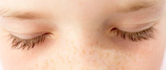
A sign of a benign tumor is the presence of growing hairs in the mole. When the nevus becomes malignant, the hairs disappear.
Removal of moles is contraindicated in cases of exacerbation of chronic diseases, unhealthy skin around the nevus and cardiovascular pathologies.
Types of moles
The concept of “good” and “bad” moles is not new for many, but what is good and what is bad can be understood without special logical conclusions.
“Good” education does not create any unpleasant sensations for a person, sometimes they even decorate his appearance. Such moles do not undergo degeneration throughout life. They have a uniform structure, fairly clear and regular boundaries, and can be in the form of a spot or a knot, protruding above the surface of the skin. Moles can vary greatly in color, ranging from yellowish skin color to brown or even black. Such formations are most often homogeneous.
“Bad” moles are capable of degeneration and can contribute to the appearance of malignant neoplasms. In such cases, skin deformation, scarring, and even disability may occur. Melanoma is one of the most malignant and fastest-growing diseases. It does not occur often, so the issue of premature diagnosis is very important.
Malignant moles differ from benign ones in that their symptoms vary.
Ways to remove moles
Depending on the indications, removal of moles and papillomas on the body, face and head can be carried out using several methods.
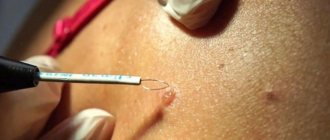
Removal of moles by electrocoagulation
A special coagulator is used to destroy birthmarks. The procedure is performed under local anesthesia. The nevus is excised with high-frequency current, which simultaneously coagulates the tissue. This helps prevent infection and bleeding of the postoperative wound. A crust forms at the site of the pigment spot, which disappears after 8-10 days. Often, after electrocoagulation, a slight scar remains. This technique is effective for combating small moles.
Mole removal with nitrogen
Cryodestruction is used to remove moles that involve the upper layers of the epidermis. Flat nevi are removed with a cotton swab, which is pre-moistened in nitrogen. Applications take no more than 2-3 minutes. During the procedure, the patient may experience minor pain and tingling. When the nevus grows deeply, a cryodestructor (a thin needle with a temperature sensor) is used. The crust that forms at the site of the mole falls off and heals within 1.5 months. In case of individual nitrogen intolerance, cryodestruction is not performed.
Laser mole removal
In this case, the destruction of birthmarks is carried out with a powerful light beam, which destroys and evaporates pathological tissue layer by layer. The procedure is bloodless and low-traumatic. The laser flow parameters are selected individually by the doctor. A crust also forms at the site of the nevus, which disappears after 1.5-2 weeks.
Radio wave removal of moles
Removing birthmarks with a radio knife is a non-traumatic, non-contact technique that involves coagulation and cutting of tissue with high-frequency radio waves. The procedure has a short recovery period and minimal complications. Radio wave excision of moles is contraindicated in cases of fever, herpes and the presence of a pacemaker.
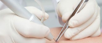
Surgical removal of a mole
The most effective method of combating hanging, large and malignant moles. The procedure involves excision of the nevus with a scalpel, followed by suturing. A scar remains at the site of the removed tumor, which heals in 3–4 weeks. A postoperative wound requires special care.
When and why to see a doctor?
Only a specialist will be able to determine whether degeneration has occurred or not. In this case, you should contact a dermatologist or dermato-oncologist, who, using a special device - a dermatoscope, will be able to examine the formation under high magnification without removing it. If any doubt remains, the doctor will be able to take a superficial biopsy and conduct additional research. You should consult a doctor in the following cases:
- if a change is noticed in the surface of the mole, for example, the skin pattern has disappeared, and a smooth spot has appeared, the skin striations have disappeared;
- if hair grew on the mole, which indicates a benign formation, and then it fell out;
- if red or blue blotches or blood vessels appear on the surface of the mole;
- if there is itching, burning, bleeding in the area of the mole;
- if additional formations and shoots appear next to the mole.
Read also Trichomoniasis - symptoms, treatment, diagnosis
What is the best way to remove moles?
The listed methods for removing skin tumors are used to completely get rid of pathological tissue. The doctor decides which method of destruction of moles to choose after a preliminary consultation, taking into account the number, location and size of age spots.
Laser mole removal is ideal for benign growths, since after their excision it is impossible to conduct a laboratory examination of the removed tissue.
The effect of cold treatment is not always predictable. The doctor may not calculate the intensity and depth of the impact, which leads to tissue burns and the formation of a postoperative scar.
The most effective methods for removing birthmarks are electrocoagulation and radio wave excision. However, they are used only for the destruction of small moles.
Despite the advantages of modern methods, in some situations only surgical removal of moles is indicated. Using this technique, you can get rid of deep, extensive formations and conduct a histological analysis of the removed tissue. Surgical excision of nevi has the longest recovery period and a fairly large number of possible postoperative complications.
How does laser removal work?
To understand how laser nevus removal occurs, you need to understand the principle of exposure to rays during the procedure. A laser wave of a certain magnitude, directed at the mole, removes the dermis layer by layer, performing excision. The high precision of the laser beam “evaporates” the contents of the mole without damaging the adjacent skin. Correctly calculated by the doctor, the beam force “seals” the vessels in contact with the operation site, which avoids bruising and inflammation.
Under laser exposure, surface skin cells begin to divide more actively, and regeneration accelerates. After a few days, the burned area where the nevus was located is completely renewed. A safe procedure for laser removal of moles takes a few minutes, without causing pain to the patient. During laser operation, a slight burning odor may be felt.
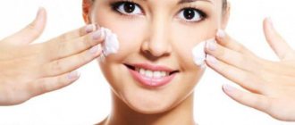
The thin red crust that appears quickly disappears if you apply a healing cream prescribed by the doctor. At the request of the patient, parts of the tumor can be submitted for histological examination to ensure that it is benign. At the end of the removal process, the area of skin on which the procedure was performed requires special care. You should not sunbathe in the sun for some time, especially if it was an open area on the neck or face.
- Treatment of dysentery in adults at home. Drug and alternative treatment of dysentery in adults
- Homemade roast pork without onions
- Potatoes with mushrooms in a slow cooker: recipes with photos
Rehabilitation period after mole removal
The speed of wound healing after mole removal will depend on the amount of tissue excised. The larger the nevus, the longer the recovery period will take. It is not recommended to rip off and touch the crust that forms on the spot after removing a mole. Over time, it disappears on its own and in its place remains a pink spot, which should be protected from exposure to sunlight. If the birthmark was removed in an area that cannot be covered with clothing, you can use sunscreen (the most optimal product will be selected by your doctor). This will avoid the risk of new moles forming. Care of a postoperative wound should be carried out until the healing area is comparable to the rest of the skin in structure and color. Since surgical removal of a mole is considered the most traumatic, it requires special postoperative care.
In case of damage, lumps or bleeding of the operated area, you should consult a doctor.
Attention!
This article is posted for informational purposes only and under no circumstances constitutes scientific material or medical advice and should not serve as a substitute for an in-person consultation with a professional physician.
For diagnostics, diagnosis and treatment, contact qualified doctors! Number of reads: 2366 Date of publication: 10/18/2017
Dermatologists - search service and appointment with dermatologists in Moscow
ABCD dermoscopic rule
In dermatology, there is the ABCD rule, which represents diagnostic signs that allow one to clearly limit a particular structure of a mole.
Read also Genital warts (condylomas) - causes, treatment
A – asymmetry. Benign formations have a symmetrical shape, that is, one half is similar to the other.
B – side, border. Benign formations have clear, even boundaries, without any irregularities, outgrowths, or processes.
C – color, color. The color of the mole should be uniform. If there are any inclusions, the appearance of red or blue shades, then you need to think that something wrong is happening to the mole.
D – formation diameter. If the diameter increases throughout life, it increases evenly.
What is the danger?
The main danger of pigmented areas of the skin is their possible degeneration into a malignant tumor - melanoma. Pigment cells are the substrate of melanoma. This is a very dangerous malignant tumor. It has no equal in its ability to quickly metastasize.
The danger also lies in some insidiousness of this neoplasm - while only a small pigment defect may be present on the skin, tumor metastases may already occur in most vital organs.
According to their oncological potential, all moles can be divided into 2 groups:
- Melanoma-dangerous (blue nevus, Ott's nevus, pigment borderline, giant pigmented, Dubreuil's melanosis).
- Melanoma-dangerous (Setton's nevus, intradermal pigmented, papillomatous, fibroepithelial, verrucous nevi, “Mongolian spot”).
Ordinary birthmarks have the following characteristics: smooth borders, small sizes, uniform coloring, almost do not rise above the skin level, do not hurt, and nothing stands out from them. Dangerous moles are characterized by the following signs:
- large formations (more than 1 cm);
- moles that change over time.
All your moles need to be closely monitored. Experts recommend photographing them at certain intervals. This way you will be able to notice even the smallest changes. There are signs that should alert you and force you to consult a specialist to determine further tactics. Some moles may need to be removed.
Signs of dangerous moles:
- the surface becomes glossy;
- change in the nature of the pattern of pigment formation;
- the usual shape of the nevus changes (its asymmetry appears, its outlines become irregular);
- the mole begins to grow horizontally (its area increases) or vertically (begins to rise above the skin level);
- the nevus begins to itch and burn;
- crusts and discharge of various types (serous fluid, blood) may appear on the surface;
- hair loss in education;
- the nevus changes its color, it becomes uneven, with alternating hypopigmentation and hyperpigmentation;
- the tissue of the birthmark thickens or becomes very soft;
- signs of inflammation in the area of pigment formation;
- the appearance of erosions, ulcers or nodules on the surface of the mole.
| Normal mole | Possible melanoma | Sign | Description |
| Asymmetry | One half of the mole does not match the other | ||
| Rough border | When the edges of a mole are uneven or irregularly shaped | ||
| Uneven color | When a mole partially changes color or the color of the surrounding skin changes | ||
| Large diameter | The diameter of the mole is more than 6mm |
What to do if a person rips off a mole
From early childhood, our parents teach us that moles should not be removed. But where did this ban come from and what is behind it? An indisputable fact is that moles must be treated with care. No one in their right mind would rip nevi off their skin. But what to do if you accidentally ripped off a mole?
It often happens that a pigment formation existed for a long time and did not cause any discomfort to a person, but after accidental trauma to its surface, it began to actively grow and change in appearance. Such symptoms are extremely dangerous, because they indicate possible malignant degeneration.
Emergency care - first you need to calm down (although this is dangerous, but not fatal), then you will have to stop the bleeding, which is often heavy and prolonged. To do this, moisten a cotton swab or bandage with hydrogen peroxide and press firmly to the wound surface, hold for about 15 minutes. As a rule, this is quite enough.
It must be remembered that it is extremely important to visit a doctor so that a specialist can determine further tactics: is it possible to remove moles or let them remain better.
Next, you need to have your mole checked. To do this, the doctor will conduct an examination and biopsy (take a microscopic piece of tissue to examine under a microscope). Such an examination will make it possible to determine whether your nevus is benign or malignant.
Only then can you delete it. The doctor will tell you about existing modern methods of getting rid of moles. If you have moles removed on your face and other areas of the skin after they have been injured, then you will not have to worry at all about how the formation will behave in the future.
If you want to get rid of pigment formation, then under no circumstances rip it off. The consequences can be catastrophic. It is better to do laser removal of moles. It's painless, fast, safe, and inexpensive.