Don't give up on beauty: facial scars can be removed, and we'll tell you how it's done.
The formation of connective tissue after various skin injuries is not just a cosmetic flaw. Scar changes can affect the functionality of moving parts of the body, increase psychological depression and manifestations of post-traumatic syndrome.
Removing scars on the face involves many different ways to influence the defect, we will tell you about them today in our article.
Why do scars form?
Scars and scars are synonymous concepts of the same word. Literally translated from German, the word “scar” is a scar, but in generally accepted terminology, a scar denotes a more scientific definition of a defect, and a scar is an everyday and commonly used term.
There are many factors that provoke the appearance of noticeable scars, but each of them is based on any violation of the integrity of the skin. It is noteworthy that they can also form inside our body, on organs during diseases and operations, but the fact that they are not visible does not reduce the consequences: the area replaced by connective tissue cells does not participate in the functions of the organ, or deforms it.
loading…
Scars on the face occur for the following reasons:
- any types of damage: burns, cuts, operations, especially if the process was accompanied by suppuration;
- chronic forms of acne;
- deep purulent skin diseases: carbuncles, boils, phlegmon, abscesses;
- features of the regenerative function of the skin, the tendency to form scars with minor damage.
The latter reason has a direct impact on the type of possible scar, its visibility, duration and effectiveness of treatment.
The following factors also influence the formation of a defect:
- Evenness of the edges of the wound (lacerations have a high risk of displacement, therefore, they scar more noticeably);
- Depth of damage. The peculiarity of connective tissue is that it has a universal composition and cannot differentiate into different types of cells. Essentially, a scar is an orderly arrangement of collagen fibers with fibroblasts that are unable to support organ function. The main purpose of connective tissue is to create a “patch” at the site of damage. Thus, with sufficiently deep damage, this patch can only either tighten the edges or, if possible, fill the voids with elastic collagen strands.
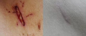
- Duration of presence on the skin. Fresh scars, with the right approach, can be significantly reduced with the help of agents that accelerate regeneration, improve blood circulation and absorb excess collagen. Old scars are denser and over time they deform the skin to suit themselves (after all, the outer layer is highly susceptible to the influence of factors such as height and weight changes).
Treatment of scars is not an easy task; various methods are used to solve it, which depend on the type of scar and the characteristics of the patient. When talking about removing scars in a particular area of the human body, it is more rational to use the term “correction”, since it is not always possible to eliminate the defect completely, but it is quite possible to reduce its severity and make it invisible to others.
Next, let's talk about different types of scars, since the method of dealing with it depends on the type of scar.
Contraindications to scar removal
Treatment of postoperative scars may have contraindications. The choice of specific technology depends on the type of scar and its maturity. Some methods are used both for the treatment of immature scars and fully formed ones (ultrasounds, mechanical-vacuum effects). Phonophoresis is not acceptable for scars younger than 3 months, and microcurrents cannot be used on old wounds.
Surgical excision in patients prone to keloid formation results in relapse in 80% of cases. It is rare that this method of therapy is recommended.
Cryotherapy is contraindicated for people with dark skin tones, as exposure to low temperatures leads to local depigmentation. Electrophoresis therapy is unacceptable in the presence of open wounds, and when treating scars that are more than a year old, this procedure is not very effective.
Each case must be reviewed by a doctor. Having assessed the type of scar, its age, area and density, the specialist will prescribe an effective method, also taking into account the patient’s allergies to the drugs used.
Normotrophic scar: description
Most often occurs with minor damage to the epidermis and shallow damage to the hypodermis.
Characteristic features of a normotrophic scar:
- does not differ from the level of healthy skin, is clearly level with it;
- the color is usually pale pink, but, due to individual skin characteristics, the shade can vary from pale red to brown;
- Over time, it can lighten and become almost invisible.
This type of scarring is considered the most physiological and most favorable for treatment.
Clinical variants of scars
Retracted, deforming (depressed) scars in the form of pits and craters are the most common types of scars after acne, chickenpox and skin tuberculosis. They are most often localized on the face and range from pitted scars to deep crater-like defects. Pit-shaped scars often merge and form wide mesh scars (Fig. 13.3). The edges of scars can be steep or flat. Steep edges cast a shadow, making them more visible. Beveled, sloping edges allow light to pass to the base of the scar without casting a shadow, so they are less noticeable.
Rice. 13.3. Deep crater-shaped scars after healing of nodulocystic acne
The histological picture of such forms of scars is variable. The unevenly thickened epidermis is pressed into the dermis in the form of a ribbon. In the dermis, channels filled with horny contents are identified. Along with inflammatory infiltrates, some of which are susceptible to fibrosis, signs of granulomatosis are present in the dermis. The severity of fibrosis and, consequently, the severity of scars depend on the stage and severity of previous inflammatory lesions.
Flat atrophic small scars occur mainly on the face. Large scars, up to several centimeters in size, are noted in the shoulder girdle area. Atrophic scars develop in patients suffering from conglobate acne, lupus erythematosus, tubercular tertiary syphilis, etc. As a rule, they are the result, on the one hand, of a normo- or hyperergic reaction of connective tissue to damage, and on the other hand, relatively favorable wound healing conditions.
Fresh scars have a wide range of colors - from pink to purple, old scars - from alabaster white to yellow. Atrophic scars are usually very thin (the thickness of tissue paper) and have a folded transparent surface through which the vessels are visible. As a result of fibrosis, all skin appendages disappear, its relief is completely smoothed (Fig. 13.4).
Rice. 13.4. Atrophic smoothed scars
Histological examination reveals a pronounced flat thin epidermis with voids. Within the dermis, a large number of dilated lymphatic and venous vessels are found. Thin, horizontally located collagen fibers in the form of nodes and loops are clearly visible; Lymphohistiocytic cells are located between these fibers. Remnants of muscles that lift the hair, nerve fibers, organic matter, giant cells, calcifications or even bone formations are also identified.
Raised perifollicular papular scars are small in size, dense, hard, slowly growing, oval in shape, slightly raised above the surface of the skin (up to 2-3 mm), white or yellowish in color. They are never found on the face, but are localized exclusively on the back and chest. Some authors describe this type of scars as papular acne scars, perifollicular elastolysis, and anetoderma-like acne scars. Such scars are often mistaken for connective tissue nevus or closed comedones. Puncture of the top of the scar with a needle allows you to distinguish them from comedones (in the latter case there will be a sebaceous discharge: Fig. 13.5).
Rice. 13.5. Perifollicular papular scars
Histological examination reveals that the perifollicular papular scar is significantly larger in size than those determined visually. The elastic fibers in it are completely destroyed and are preserved only along the periphery of the scar. The skin appendages are also completely destroyed, only sometimes remnants of hair follicles are found.
Hypertrophic scars are dense tumor-like formations protruding above the level of the surrounding skin with a moderately or slightly lumpy shiny surface, sometimes covered with flaky epidermis. At first they are bright red in color, later becoming yellow-white. Most often, scars are located on the back, shoulders or sternum. Often, transverse cracks form in places of tension in the scar, and areas exposed to pressure or friction may ulcerate. Densified scars without clear boundaries become atrophic and never spread beyond the damaged area. Their shape and size are varied - from narrow cord-like cords and membranous folds to extensive scar areas (Fig. 13.6). Hypertrophic scars form within 6-12 months after epithelization of wounds that heal by secondary intention, with the formation of excessive granulations in them, followed by gross fibrosis. Therefore, such scars are also called fibrous nodes.
Rice. 13.6. Hypertrophic scar
In the formation of hypertrophic scars, the large size of the wound defect and constant traumatization of the scar play an important role. Trauma is more pronounced in functionally active areas, especially if the scars are located parallel to the direction of muscle contraction.
On scars located transversely or obliquely to the direction of muscle contraction, movements do not have any significant effect. Constant irritation, ulcerations and ulcerations of the epidermis support a chronic inflammatory process that prevents the softening of the scar. Over time (after years and even decades), the scars become flattened, gradually acquiring clear outlines, delimited from the atrophic part of the scar and intact skin. They are often accompanied by itching.
Histological studies of hypertrophic scars show that they consist exclusively of randomly arranged dense collagen fibers of various sizes, most of which are located in the horizontal plane. The skin appendages are completely destroyed. Elastic fibers are either absent or rare. The vascular network is very sparse (a picture of pronounced fibrosis). Insufficient maturation of scar tissue is noted, i.e., the presence of granulation residues, the presence of myofibroblasts, and a large number of cells, which distinguishes hypertrophic scars from keloid scars, characterized by excessive accumulation of the extracellular matrix. The presence of macrophages and fibroblasts in these scars determines the high level of TGF-β - 3 times higher than in a normal scar. TGF-β is the most powerful stimulator of collagen synthesis and the only known inducer of the transformation of fibroblasts into myofibroblasts. Therefore, hypertrophic scars, characterized by increased fibrosis, are subject to active retraction.
Calcified scars . Sometimes an inflammatory process in tuberculosis. syphilis, globular acne, scleroderma ends with the formation of osteomas or calcifications in the scar area. These formations are diagnosed clinically. They can form on the face, especially in the eyelids, forehead, and upper back. On x-rays taken for various diseases, calcified scars are clearly projected in the form of scattered multiple shadows, often interpreted as artifacts. In some cases, calcified scars lead to facial disfigurement. Upon palpation in such patients, large nodes of stony density are determined.
Keloid scars are a special, most severe group of scars that differ from others in appearance and pathogenesis. A keloid scar is a dense, tuberous tumor-like growth of connective tissue, most often located in the sternum, on the lateral surfaces of the upper extremities, shoulders, back, neck, in the area of the body of the lower jaw, and the nasolabial triangle. The color of scars varies from dark red to brown with a complete absence of skin microrelief and pores. Thick, shiny, sharply raised lobular fibrokeloid plaques sit tightly on the skin and have a strong tendency to recur after removal. Old scars become even more dense and cyanotic or hyperpigmented.
The development of keloid tissue occurs according to the embryonic type, i.e. loss of the initial stage of collagen formation is observed, due to which the scarring process becomes pathological. The main role in the formation of keloid is assigned to a violation of the relationship between the breakdown and synthesis of collagen in the direction of increasing the latter. The cycle of changes is repeated many times, and the balance between the formation of cells, the number of vessels, structures and the synthesis of the main substance of connective tissue differs from the ratio inherent in a regular scar. As a rule, the appearance of keloids is preceded by skin damage, sometimes unidentified (Fig. 13.7). With acne, keloid scars can follow inflammatory eruptions, even simple papulopustular ones, but most often they are provoked by globular acne.
13.7. Keloid scar developed after injection
The development of keloid scars begins several weeks or months after wound healing, when limited compactions appear in the thickness of the scar tissue, rapidly increasing with the involvement of visually unchanged surrounding tissues in the process. This process lasts from several months to several years and is accompanied by itching, which is probably due to the presence of a large number of mast cells. Sometimes sensory disturbances (dysesthesia) or pain are noted, especially in patients who have scars after thoracotomy and sternotomy. Keloid scars are capable of penetrating into the subcutaneous tissue, but very rarely ulcerate. Scars located on the earlobes tend to form “petioles,” while when localized in the upper half of the body, they are wide, raised, often claw-shaped, and are more common, rapidly growing, and resistant to therapy. Keloid scars are often mistaken for hypertrophic scars, and vice versa.
Below are clinical signs that help differentiate hypertrophic and keloid scars.
Histological studies show that keloid scars are covered with an even layer of epidermis without protrusions into the underlying layers of the dermis. The thickened epidermis along the entire length of the scar retains approximately the same structure of all layers, thickens at the edges and grows in the form of acanthosis, but never exfoliates or flakes off. With a true keloid scar, in contrast to a hypertrophic scar, a “bed-like network” and “skin papillae” are microscopically determined, since scar formation occurs in the absence of tissue deficiency. With hypertrophy, this deficiency is compensated by scar tissue, so these formations are absent and the scar does not spread over the surface beyond the initial damage; The keloid scar penetrates into healthy tissues, going beyond the destroyed ones. Clinically, the type of scar is assessed visually and by palpation. In addition, with keloid scars, areas of active synthesis and degeneration (hyalinosis) of collagen fibers are identified. Collagen fibers are disoriented, have a spiral, nodular or bundled organization.
Morphologically, collagen fibers consist of immature forms of collagen with a low degree of orientational ordering of its macromolecules. The cells are dominated by young fibroblasts with high activity, numerous mast cells and macrophages. Among the fractions of glycosaminoglycans, sulfated ones dominate. In tissues, swelling of the intercellular substance, vasodilation, stasis phenomena, erythrodiapelosis, and desquamation of the vascular endothelium are noted. All this reflects pathological tissue regeneration, which occurs under conditions of inflammation and hypoxia and is accompanied by impaired differentiation of scar tissue.
Hypertrophic scar: characteristics
The prefix “hyper” is used in medicine to indicate excess. Regarding scars, she says that the process of skin restoration was accompanied by excessive formation of collagen fibers.
Therefore, a hypertrophic scar can be distinguished by the following features:
- it always rises significantly above the surface of healthy skin;
- does not protrude beyond the edges of the wound area, that is, it grows upward, but not in breadth;
- such a scar is very noticeable on the skin of the face because it has a dark color: crimson or deep purple;
Over time, the hypertrophic scar may become less noticeable, provided that some of the collagen from the connective tissue is absorbed.
Atrophic scar: how to recognize
This scar, unlike the previous one, develops due to insufficient production of collagen fibers and, therefore, insufficient filling of voids in the damaged skin.
Therefore, an atrophic scar has the following features:
- always below the level of healthy skin;
- has a paler color, almost white, compared to other areas;
- accompanied by impaired blood microcirculation in this area;
- Externally, the scar is covered with fairly thin connective tissue, which makes it easily injured.
A special type of atrophic scar is stretch marks (scientifically, stretch marks) - the only type of scars that occurs without external damage to the skin, but due to internal rupture of collagen fibers with subsequent improper fusion and scar formation according to the atrophic principle.
Stretch marks on the face can occur in the cheeks, forehead and under the chin with sudden fluctuations in body weight, from hormonal imbalance, after pregnancy, but can be associated with skin diseases accompanied by pathology of collagen formation: Marfan syndrome, Ehlers-Danlos syndrome. You can learn more about scars of this type and methods of treating them in the article “ Treatment of atrophic scars on the face .
Treatment at home
It is recommended to treat hypotrophic scars at home only on immature and shallow formations. But even in this case, the result is not always pleasing.
Folk remedies
To smooth out and lighten the defect a little, try making a clay mask.
You will need:
- white or green clay - 50 g;
- lemon juice - 50 ml;
- tea tree oil - 2-3 drops.
Mix the ingredients thoroughly and apply locally to the affected area. After 30 minutes, wash your face.
A mixture of mumiyo and baby cream smooths out linear scars well. Apply the application to the defect for 40 minutes, then apply pharmaceutical ointment.
If you have real beeswax at home, melt it in a double boiler and mix with olive oil. Place the warm mixture on gauze and apply to the scar.
When using folk remedies to treat atrophic scars on the face, remember that they are not effective enough and do not expect miracles. Judging by the reviews, home treatment can only slightly smooth out and lighten the scar tissue.
Pharmacy drugs
Pharmacy creams are more effective for removing blemishes on the face. True, they only help on immature scars.
The most effective drugs:
- collagen Emalan-gel. Judging by customer reviews, this is the best remedy for scars. The price of the bottle is 600–700 rubles;
- Kelofibrase. The main component of the drug is urea, which moisturizes, softens and activates blood circulation at the site of the lesion;
- Contractubex. Quite expensive, but effective. The cost of a 50 ml tube is 800–900 rubles;
- Dermatix-gel. New generation drug. It copes well with scars, but is also not cheap - 2000–2300 rubles.
Dermatologists believe that pharmaceutical ointments cannot get rid of atrophic scars when used independently. It is much more effective to use them as an addition to hardware procedures.
Cosmetic mud
Cosmetic mud applications are considered an effective way to eliminate scarring on the face. The healing effect is often present even in cases where folk remedies and ointments are powerless. Mud perfectly softens connective tissue, activates collagen synthesis, and reduces the depth of the lesion.
To treat hypotrophic scars on the face, it is recommended to use Saki mud. Based on them, many effective preparations have been made for removing scars, stretch marks and other skin lesions.
The most popular of them:
- silt mud “Formula of Health”, doy-pack 1 kg - 240 rubles;
- Saki mud “Crimean natural collection”, 700 gr. — 230 rub.;
- applications with mud from Lake Saki - 1,500 rubles;
- silt sulfide mud “Manufactory House of Nature”, 1200 gr. — 320 rub.
Therapeutic applications have an anti-inflammatory and absorbable effect, improve breathing and tissue trophism, so they can be used both at the beginning of the regeneration process and on mature scars.
Keloid: what is it and why does it occur?
Keloid scars are the most insidious and difficult to treat. They are formed, as a rule, due to a genetic predisposition to this type of scarring of lesions.
These scars look like this:
- Convex, knotty, purple.
- They resemble a hypertrophic scar, but over time they can enlarge and involve healthy areas.
- All of these types of scars are usually painless, but keloid scars hurt and itch even in their mature state.
- Such a scar can occur even with minor damage to the skin.
Keloids can reoccur after correction. But they are the ones who especially need regular treatment, since in addition to aesthetic discomfort they cause physical suffering.
Methods for dealing with facial scars
The method of getting rid of a scar depends on the following characteristics of the defect:
- degree of prescription of formation;
- types of scar;
- depth of damage
- associated complications: severe deformation of the injured area, contracture (limitation of motor function if the scar tightens moving areas, on the face, for example, the eyelids and lip area, on the body - the inner surface of the joints);
- individual features of restoration of damaged structures
- type of damage: linear cuts or stitches are easier to make less noticeable than extensive burn scars.
Given these characteristics, different types of treatment can be applied. We have prepared characteristics of each of them for you.
Features of the procedure
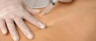
There are two types of mesotherapy: injection and non-injection, each of which is used in certain situations. Injection mesotherapy of the face and other areas is performed using thin needles or using a special mesoroller. The procedure begins with cleansing the skin, then applying an anesthetic cream. The number of injections depends on the area of the defect and is calculated individually. The non-injection method is performed without puncturing tissue. The active substances are delivered to the cells using ultrasonic waves and low frequency current. The procedure begins with cleaning and disinfecting the skin, after which a pre-selected preparation is applied to the problem area. At the same time, this area of the skin is exposed to ultrasound or another type of wave.
Early conservative therapy with medications
It is used at the stage of formation of connective tissue after primary epithelization of the damaged area. It is necessary to soften the fabric and give it elasticity.
For this purpose, special absorbable gels are prescribed:
- Contractubex;
- Mederma;
- Kelofibrase;
- Fermenkol;
- Kelo-cote.
If there are signs of connective tissue hypertrophy, a good effect is shown by applying an occlusive dressing to the area of scarring - pressotherapy. For these purposes, special silicone patches are also used, which, when worn for a long time, help smooth out the developing scar.
Let us remind you that these methods can only be effective when working with young scars; the appropriateness of their use is determined by a specialist of the appropriate medical profile.
Peeling for facial scars
To solve the problem, chemical medium and deep peeling, combined Jessner peeling, and microdermabrasion are used. Such methods are effective in combating the manifestations of post-acne and hypertrophic scars.
During the process of exposure to peeling substances, the top layer of connective tissue is destroyed, while skin cell renewal is stimulated, which allows normalizing collagen production and adding elasticity.
Dermabrasion and microdermabrasion of the skin are based on the same principle, in which mechanical exfoliation of the upper layer of skin occurs using a stiff brush or hard microparticles. Then the epidermis is restored and renewed with improved characteristics, which helps visually reduce the severity of the defect.
To achieve a positive effect, it is necessary to conduct several sessions, as well as follow all recommendations for skin care after the procedure. It should also be remembered that professional peelings are not recommended during periods of solar activity: from spring to late autumn.
How to care for your skin after mesotherapy
After the procedure, the skin needs proper care, which involves following a few simple rules:
- Avoid exposure to ultraviolet and sunlight, use special creams to prevent pigmentation.
- Refuse to use cosmetics.
- Reduce physical activity.
- To refuse from bad habits.
- Limit visits to the sauna, solarium, and swimming pool.
An exact list of recommendations is compiled individually for each patient, taking into account the characteristics of the mesotherapy performed.
Injection method to combat scars
Depending on the type of scar, the following methods of eliminating them using injections are used:
- Atrophic scars , first of all, need to be raised and leveled with the general level of the dermis. Therefore, it is recommended to fill them with filler or collagen by injection. The disadvantage of this method is contact injury to the scar. And over time, the injected drugs dissolve and the defect becomes noticeable again.
- Keloids and hypertrophic scars , on the contrary, require the introduction of special absorbable agents; for this, Diprospan is injected into the base of the scar. This helps to reduce the activity of formation of collagen threads, ease their tension, that is, quickly reduce the pain of the keloid.
Mesotherapy practice has similar properties. In this case, the introduction of special meso-cocktails with hyaluronic acid through micro-punctures of the skin is used. Also, in the early stages of the formation of skin changes on the face, a mesoscooter and special serums can be used.
In some cases, plasma lifeling is an effective remedy: injection of autoplasma enriched with platelets.
Physiotherapy against scars
This method involves the hardware introduction of absorbable agents into the middle layers of the skin, which helps to significantly soften scar tissue.
The following physiotherapeutic techniques are used:
- iontophoresis with lidase, collagenase;
- phonophoresis (ultrasound effect on the scar) with corticosteroid or absorbable ointments;
- shock wave therapy - mechanical destruction of pathological tissue;
Such methods help to cope with the pain associated with the scar, as well as significantly reduce inflammation, if present.
Laser scar removal
The laser is used to treat scars of any type, not only on the face, but also on the body. This method allows you to reduce the severity of the pathological defect in a fairly short time.
For this purpose, the following laser treatment methods are used:
- fractional laser resurfacing (photothermolysis);
- CO2 laser polishing;
- erbium laser;
- exposure using pulsed light radiation.
With this method, non-contact destruction of scar tissue occurs using a hot beam, which has the ability to penetrate the skin to different depths, healthy areas are not affected.
Laser radiation most effectively and atraumatically eliminates pathological fibroblasts and destroys dense scar collagen, and also powerfully stimulates the growth of healthy skin cells.
Thanks to this technology, in a fairly short time you will be able to evaluate the result before and after the procedure. But this may require several sessions (on average from 2 to 10 depending on the characteristics of the defect).
What determines the effectiveness of mesotherapy and PRP in the treatment of post-acne
Mesotherapy is a percutaneous non-ablative collagen-inducing therapy. During the procedure, thousands of microchannels are created on the patient's skin using a mesoscooter or an electronic device, the depth of which reaches the papillary layer of the dermis.
Activation of fibroblasts in response to skin damage with subsequent release of growth factors and cytokines, as well as changes in the electrical potential of the epidermis/dermis, cause physiological effects - neocollagenesis and neoangiogenesis. The accumulation of collagen in areas of atrophic scars promotes tissue remodeling and smoothing of the skin. The platelet concentration in PRP, which is obtained by centrifuging the patient’s own blood, is over 1x10⁶ units/μl.
After the administration of platelet-rich plasma, as platelets are activated, growth factors begin to be released, promoting the activation of fibroblasts and the synthesis of new collagen:
• platelet-derived growth factor;
• transforming growth factor;
• insulin-like growth factor.
Thus, the complementary effect of mesotherapy and PRP is due to the healing mechanisms that are triggered in response to skin damage and the action of the main growth factors involved in this process.
Facial mesotherapy: what are the side effects and how to avoid them
Cryodestruction: is it possible to freeze a scar?
Cold treatment of the scar is used for keloid and hypertrophic scars. The purpose of this procedure is to eliminate as much as possible the protrusion of a pathological formation on the skin. Using an applicator, liquid nitrogen is applied to the area, which destroys excess collagen, forming a blister like a burn.
Over time, healthy tissue begins to form under the crust, which contributes to the resorption of the scar. The procedure is performed under local anesthesia. The effect can be assessed when a relatively healthy covering is formed at the site of the scar and the skin tone is evened out. On average, this period is up to six months.
In some cases, a combination of cryodestruction and Bucca irradiation is effective - exposure of the scar to X-ray radiation with a load that is safe for the body. This method is used to treat old keloids.
Surgical method of removal
This method of treatment is most often used in relation to keloid scars, because they tend to reappear over time.
There are several ways to deal with scars using surgery:
- Z-plasty: a special type of incision that helps guide the edges of the scar in accordance with the natural lines of the skin;
- skin transplantation and skin grafting - transplantation of a flap from a healthy area to a scarred surface;
- an alternative multi-stage method: a special silicone reservoir is introduced under the area of overgrown connective tissue, which is periodically fed with saline through a puncture. The sac gradually increases in size, stretching the skin, while forming healthy layers of tissue. When the area of new healthy areas around the scar has grown sufficiently, the reservoir is removed, the pathological tissue is excised, and the formed edges of the new skin, which appeared as a result of stretching, are carefully sewn together with a cosmetic suture.
Each method of surgical intervention may be accompanied by certain risks, such as increased scarring and secondary infection, so it must be used strictly according to indications.
Indications and contraindications
Mesotherapy is an effective way to treat many cosmetic defects.
In addition to treating post-acne (scars, spots), it can give a noticeable positive result in the following cases:
- signs of premature skin aging, the appearance of characteristic facial wrinkles;
- darkening of the skin as a result of previous diseases, the consequences of hormonal transition periods (adolescence, pregnancy, menopause), aggressive treatment of acne;
- the appearance of dark circles under the eyes, age spots, freckles, vascular diseases in which the capillary networks become visible to the naked eye;
- scars of any origin: surgery, post-acne, atrophic manifestations of the skin, keloids.
Mesotherapy for acne scars can help even in cases where previous long-term treatment led to complications and new inflammatory processes began to appear directly in the scars themselves.
This cosmetic procedure is strictly contraindicated in the following cases:
- during any trimester of pregnancy and breastfeeding, since the active ingredients used can have a negative effect on the body of the fetus or newborn;
- persons under 18 years of age;
- neoplasms of a benign or oncological nature;
- cholelithiasis;
- chronic pathologies of the urinary organs;
- period of exacerbation of chronic diseases of the gastrointestinal tract, cardiovascular system;
- the appearance of new inflammations on the skin;
- the period after infectious inflammatory diseases of a bacterial or viral nature;
- allergic reaction to the drugs used, individual intolerance;
- blood clotting pathologies;
- small cracks, wounds, injuries, various damage to the skin.
Before carrying out the procedure, a dermatologist must conduct a full examination and questioning of the patient, as well as allergy tests in order to exclude any possibility of the development of unforeseen situations and complications.
What methods are suitable for a child?
You can get injuries that will later remind you of themselves by forming a scar at absolutely any age, but it is precisely the period of childhood that is most conducive to this. The characteristics of children's skin are such that it grows rapidly, because the baby himself is growing.
And scar tissue may not keep up with this growth due to the fact that scar collagen is permeated with connective tissue cells and is less elastic. This poses a great danger as it can lead to facial deformation.
On the other hand, if the damage was shallow and the child has not yet reached the age of eight, there is a high chance that the damaged area will be fully restored and no traces of the incident will be visible.
In children, it is quite difficult to predict how the scar will manifest itself after a certain time. Therefore, monitoring his condition and dynamic observation is necessary. All methods that can be aimed at reducing the severity of the consequences of injury should be prescribed by a doctor based on life history, the state of the degree of epithelization of the wound, and the presence of complications.
To treat scars on the face, the child is initially prescribed external drug therapy. The use of silicone patches Mepiform, Scarfix, Mepiderm Arilis has proven itself well. They help ensure that the scar is sufficiently hydrated, which prevents its edges from tightening and, consequently, possible deformation.
The use of absorbable anti-scar gels is possible in the absence of an allergic reaction to the substances in the composition of the product. This type of therapy can be used at home, but only after prior consultation with a pediatric dermatologist and as prescribed.
Cosmetic procedures are also successfully used in children:
- Jessner peel;
- TCA chemical peel;
- laser resurfacing.
But it is worth considering that any type of manipulation may have limitations depending on age, skin condition, type and depth of the scar. Treatment of scar changes on the face of a child involves the most gentle intervention, therefore it is carried out in stages and requires strict control over the restoration of the skin.
Thus, involuntary tearing off of the scab formed after the procedure can lead to a worsening of the scar.
Radical surgical intervention is performed in cases where scar formation can lead to disruption of important functions: chewing, blinking, breathing (if deformation of the nose occurs).
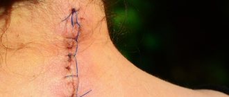
Rules for the treatment of postoperative scars
The appearance of a postoperative scar during any surgical therapy (mastectomy, cesarean section, thyroidectomy, appendicitis and others) is inevitable. To prevent the proliferation of connective tissue, silicone plates, patches, absorbable gels and ointments should be used during scar formation. It is very important to ensure that the problem area heals calmly: to get rid of rubbing, stretching, and pressure. If the patient wishes, physiotherapeutic procedures can be prescribed. Large scars on the leg or body may require further surgery.
Traditional medicine offers many recipes for getting rid of scars at home, but treatment of extensive post-burn marks and consequences of injuries (including on the forehead and lips) is best left to specialists.
The article has been verified by the editors