To the reference book Operations in otolaryngology are usually aimed at restoring hearing and may involve the installation of an auditory prosthesis.
- Operations
- Preparation
- stapedoplasty using modern prostheses;
- myringoplasty;
- tympanoplasty (hearing-improving surgery) using modern prostheses;
- general cavity (radical) surgery on the middle ear;
- mastoidoplasty;
- shunting and drainage of the tympanic cavity for exudative otitis media;
- removal of exostoses of the external auditory canal;
- revision of the tympanic cavity;
- reoperations: tympanoplasty, stapedoplasty;
- operations for tinnitus;
- operations for ear injuries;
- formation of the external auditory canal in congenital and acquired atresia.
Operations in otolaryngology are usually associated with diseases accompanied by hearing loss and requiring surgical treatment.
Indications for surgery
The operation is indicated for damage to the hearing system, which is expressed in hearing loss (of varying degrees, up to its complete loss) due to such consequences of otitis media as:
- cholesteatoma (tumor-like formation of dead epithelial cells and other substances surrounded by a capsule of connective tissue);
- perforation of the eardrum (large ruptures and extensive perforations);
- tympanosclerosis (overgrowth of connective tissue in the mucous membrane of the middle ear);
- disturbance in the chain of auditory ossicles;
- atelectasis of the tympanic cavity (strong retraction of the eardrum), etc.
Also, the cause of poor hearing and an indication for surgery may be congenital anomalies of the middle ear and mechanical damage to the eardrum (for example, due to careless removal of foreign objects from the ear).
Patient management after surgery
Postoperative treatment depends mainly on the type of intervention: all operations on the middle ear (anthrotomy, anthromastoidotomy) are characterized by an open wound, which is packed and then covered with a sterile bandage. As a rule, by the evening the patient begins to feel much better: the temperature drops, the pain goes away. The next day, the first dressing is performed; The drainage tampons are replaced with new ones, the postoperative cavity is washed and disinfected with antiseptic drugs. The following dressings are performed every few days and are stopped only after the cavity is completely filled with granulating tissues. If suppuration stops and the perforation closes, secondary sutures are not applied. In most cases, the eardrum is restored and hearing returns to normal. Purulent otitis media with a closed wound requires a general cavity sanitizing operation, in which only the external dressing should be changed and the sutures should be treated with iodine. Complete dressing is carried out only after a week. All this time, the patient is administered analgesic and antipyretic drugs.
Examination and preparation for surgery
Otoscopy
To prescribe surgery (or identify contraindications), an examination of the affected ear is prescribed, including:
- otoscopy – examination of the ear using special instruments;
- X-ray;
- bacteriological examination;
- audiometry - measuring hearing acuity using an audiometer (or tuning forks);
- passing all necessary tests.
In the preoperative period, the doctor prescribes general restorative therapy, medicinal sanitation (cleaning) of the middle ear, and other measures to improve ventilation and auditory function of the Eustachian tube.
Types of tympanoplasty
There are several types of such operations (classification according to G. Wulshtein), the choice of one of which is influenced by the nature of the damage to the ear structures:
Hearing restoration surgery
- Type I The indication for such an operation is the presence of perforation in the eardrum, but the chain of auditory ossicles must not be damaged and the patency of the auditory (Eustachian) tube must be maintained. This is the most common and simplest type of surgery. The defect is eliminated using the body's own tissues. To do this, the eardrum is lifted, a skin flap is placed over the hole and fixed on both sides using a special absorbable material. The effectiveness of this operation is about 95%. If necessary, repeated intervention may be performed;
- II type. This type of tympanoplasty is indicated for damage to such parts of the auditory ossicles of the middle ear as the manubrium, neck or head of the malleus, but the incus-stapedial joint must be intact. Plastic surgery involves prosthetics of damaged parts of the auditory ossicles;
- III type. Such plastic surgery is used in the absence of the eardrum, malleus and incus, but the stapes must be preserved. This operation does not involve the use of prostheses. Bone tissue from the body is used to restore the structures of the middle ear;
- IV type. It is advisable to perform such plastic surgery if all the auditory ossicles are missing, but the base of the stapes is preserved. In this case, the cochlear window is shielded using the preserved part of the eardrum or using a free flap;
- V type It is carried out in the case of a complete absence of all sound-conducting elements and involves fenestration of the semicircular canal (creation of a new opening in the bony labyrinth of the inner ear). Today, this type of plastic surgery is rarely performed; it has been almost completely replaced by stapedectomy (removal of the stapes and its replacement with a prosthesis).
If it is impossible to restore the eardrum and auditory ossicles at the same time, the operation is carried out in stages. First, reconstruction of the auditory ossicles (ossiculoplasty), and then plastic surgery of the eardrum (myringoplasty).
Depending on the type of operation, either local anesthesia or general anesthesia is used.
Preparation and progress of surgery
Before the operation, a diagnostic study is performed, which includes otoscopy, tympanometry and audiometry. Their data allows you to assess the extent of damage, as well as detect possible contraindications. Other ear, nose and throat conditions should be eliminated before surgery. For example, if a patient suffers from adenoiditis, then an adenoidectomy is first performed. If otitis is present, the doctor will prescribe the necessary medications to treat it, and only after the infection has been eliminated can tympanoplasty of the ear be performed.
For small perforations, tympanoplasty can be performed under local anesthesia through the ear canal, which is slightly incised and the eardrum is raised. If the perforation is very large or is in an inconvenient location, access to the tympanic cavity is carried out through an incision behind the ear. In this case, general anesthesia is needed.
The eardrum is carefully examined under a microscope, the hole in the eardrum is cleaned, and any abnormal areas are removed. If necessary, at this time reconstruction of the bones of the middle ear (ossiculoplasty) or removal of cholesteatoma from the walls of the middle ear is performed.
Interesting to know! The need for ossiculoplasty is not always known before surgery. In some cases, this only becomes apparent when the ear is completely opened and examined under a microscope.
A situation often occurs when the cartilage that connects the auditory ossicles to each other is destroyed. For their reconstruction, a small cartilage taken from the tragus of the patient's ear can be used. If the damage to the bone is too great, it is removed and a prosthesis is installed in its place, or a connection is created without it.
If there are no pathologies or damage, then the remaining operation concentrates on restoring the defect of the eardrum. To close the tear in the membrane, a piece of tissue called a graft is implanted. The eardrum is lifted and the prepared flap is inserted behind it so that it covers the perforation. A tampon is placed behind the membrane, and in order for the graft to stay in place, an absorbent gelatin sponge is placed under it (it dissolves itself after a while).
Grafts for eardrum tympanoplasty and ossiculoplasty are usually taken from a vein, fascia (muscular membrane) behind the ear, or tragus cartilage. Synthetic materials can also be used, for example, in cases where the patient has already had surgery. Finally, the incision is sutured and a bandage is placed on the ear.
Possible complications
Infection may cause pain
Possible complications after tympanoplasty include:
- infection accompanied by pain, swelling, discharge from the ear;
- no improvement in hearing;
- damage to bones and nerves, leading to various pathologies;
- displacement, atrophy or necrosis of the graft (if used);
- dizziness, imbalance;
- short-term paralysis of half the face.
In 3% of cases after tympanoplasty, hearing continues to decline until it is completely lost. Most often this occurs after non-radical tympanoplasty or complicated surgery.
Plastic surgery of the ears
Aesthetic proportions of the outer ear
The long axis of the ear, the one that runs from the upper edge of the helix through the earlobe, should be parallel to a line passing through the bridge of the nose (a) .
The points of attachment of the helix (b) and earlobe (c) to the head are called “ otobazion ”, and form a line (d) passing immediately behind the mandibular joint. The angle between lines (a) and (d) is approximately 20 degrees .
The place of attachment of the helix to the head is the superior otobasion (b) , located on the same level with the external canthal ligament of the eye (e) , the distance between these anatomical formations (b) and (e) is 65-70 mm .
The upper edge of the curl is flush with the arch of the eyebrow; these points form a line (g) . If the curl of the auricle is located above the line of the eyebrows, then such an ear can be considered large.
The lower edge of the earlobe is level with the base of the tip of the nose, they form a line (h) . If the lobe is located below the line running through the base of the tip of the nose, then we can assume that its size is larger than average - there may be a uniform increase in the entire auricle.
Analysis of the curves of the outer ear begins with the helix and antihelix. They begin from below, at the level of the tragus and diverge upward, where they are separated by the scaphoid fossa.
At the top, the antihelix is divided into a smoother, wider upper stalk and a lower stalk. When viewed from the front, the helix forms the most lateral deviation of the ear and should be only slightly visible behind the antihelix and superior crus.
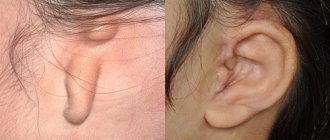
Classification of protruding ears (protruding ears)
Correct analysis of ear protrusion is an important step in otoplasty. Protruding ears (protruding ears) are the result of one or more developmental features of the ear.
- Underdevelopment of the antihelix.
- Hypertrophy of the auricle or deformation of the shape of the auricle.
- Protrusion of the lobe.
- Uniform enlargement of the entire outer ear (macrotia), or excessively rapid growth of the ear.
1 - The most common cause of protruding ears is underdevelopment of the antihelix . The spectrum of underdevelopment of the antihelix is quite wide, from its complete absence, in this case the entire auricle protrudes, to partial underdevelopment, only the upper pole of the ear occupies a protruding position.
2 - Excessive development of the cartilaginous structure of the auricle itself also leads to protrusion of the ear. It has been established that the thinner cartilages of the helix and antihelix complex are located on the stronger cartilage of the auricle and when it increases, excessive protrusion of the entire ear occurs.
3 - Protrusion of the lobe may be the only deformity of an otherwise normal ear. This may be a consequence of the unusual shape of the tail of the helix, or develop as a result of hypertrophy of the auricle.
4 - Uniform enlargement of the entire outer ear (macrotia) . The range of “normal” ear sizes is quite wide, but there are cases where increased ear sizes make it disproportionate to the facial skeleton. This may occur as a result of isolated congenital overgrowth of the ear, or in combination with faster growth of one side of the face. Uniform enlargement of the entire ear may be a sign of previous neurofibromatosis or a vascular abnormality.
Contraindications
Like any surgical intervention, tympanoplasty has contraindications. This operation cannot be performed if:
- exacerbation of chronic inflammatory processes in the middle ear;
- damage to the inner ear (labyrinth);
- intracranial or septicopyemic complications of ear diseases;
- significant damage to sound-conducting structures;
- impaired patency of the Eustachian (auditory) tube;
- general severe conditions.
If the second ear cannot hear at all, then the possibility of tympanoplasty is discussed individually, and the pros and cons are weighed.
Diagnosis of ear malformations
To diagnose ear malformations, clinical and audiometric examinations, as well as radiological methods, are used. Accurate anatomical description of malformations using imaging techniques is indispensable, especially for planning, outcome of surgical middle ear reconstructions and cochlear implantation (CI).
Clinical examinations
Newborns with ear deformities should undergo a detailed examination of the craniofacial structures. A thorough examination of the skull, face and neck is necessary for configuration, symmetry, facial proportions, masticatory apparatus, occlusion, hair and skin condition, sensory functions, speech, voice and swallowing. The function of the middle ear is studied especially carefully, since the development of the outer ear closely correlates with the development of the middle ear. Parotid fistulas or appendages, as well as facial nerve paresis/palsy may accompany ear abnormalities.
In addition to the basic examination of the ears (inspection, palpation, photographic documentation), attention is paid to any anatomical features that may increase the risk or jeopardize the success of middle ear surgery. These features include dysfunction of the auditory tube due to hypertrophy of the adenoids, severe curvature of the nasal septum, as well as the presence of a cleft palate (and submucosa).
Ear deficiencies may be associated with syndromes; therefore, changes in the internal organs (eg, heart and kidneys), nervous system, and skeleton (eg, cervical spine) must be excluded by a multidisciplinary team that includes a pediatrician, neurologist, ophthalmologist, and orthopedist. Preoperative assessment of facial nerve function is mandatory if reconstructive middle ear surgery is planned.
Audiometry
Audiometry is the most important functional test in patients with ear problems. Severe defects of the external ear, such as congenital aural atresia, often occur in combination with severe disorders of the middle ear and can affect all its structures. In such cases, a conductive hearing loss of 45-60 dB occurs, and complete conductive blockade can often be found down to a level of approximately 60 dB.
In cases of unilateral congenital aural atresia, early hearing testing in the apparently normal contralateral ear is important to identify and rule out bilateral hearing loss. Depending on the degree of functional impairment, bilateral hearing loss can significantly interfere with speech development. Thus, early rehabilitation is mandatory (hearing aids first, surgical correction if necessary).
Read also: Wax plug in the ear: symptoms, removal at home
Audiometry is possible even in an infant. Physiological studies include tympanometry (impedance measurements), otoacoustic emissions (OAE), and auditory evoked potentials (auditory brainstem responses). To establish specific threshold values, children around 3 years of age are subject to reflex and behavioral audiometric examinations, tympanometry, measurement of OAE and auditory brainstem response.
Objective measurement methods (OAE and auditory brainstem response) provide reliable results. In older children, reproducible results can be achieved by testing the auditory response using traditional pure-tone audiometry or behavioral audiometry. For accuracy, the audiological examination is repeated, especially in young children and in patients with multiple defects.
Vestibulological examination has differential diagnostic value. Violation of vestibular function does not exclude the presence of hearing.
Visualization methods
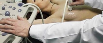
Traditional radiography is of little value in diagnosing ear defects. High-resolution computer tomography (HRCT), with its clear depiction of bony structures, is adequate to show changes in the external ear, external auditory canal (EAC), middle ear, and mastoid process. Magnetic resonance imaging (MRI) is best for imaging the membranous labyrinth, the neural structures of the internal auditory canal, and the cerebellopontine angle. HRCT and MRI are used in combination. Ultrasound diagnosis has no value for ear defects.
- CT scan.
HRCT of the temporal bone using a bone algorithm and slice thicknesses of 0.5–1 mm is suitable for assessing middle ear deficiencies. The traditional view is the axial plane, which shows both temporal bones and allows comparison of the two sides. Coronary scans are a useful important addition. Helical scanning technologies provide high spatial resolution without loss of quality and make it possible to document anatomical structures, visible variations and congenital or acquired deformities. Modern CT scans allow the reconstruction of secondary sections at any desired level in any plane, as well as the creation of three-dimensional structures.
HRCT visualizes the extent of the pneumatic cell system and the location of the jugular bulb, sigmoid sinus, and internal carotid artery. Moreover, HRCT shows the chain of auditory ossicles, the course of the tympanic and mastoid segments of the facial nerve, as well as the width of the internal auditory canal. The clear contrast between bone and air, as well as the high spatial resolution, makes this diagnostic procedure ideal for the middle ear.
Fixation of the ossicular chain cannot always be detected using HRCT. This may sometimes explain normal CT scans in patients with conductive hearing loss. The stapes is not always identified due to its small size; only sections of no more than 0.5 mm can show the entire stapes. Bone thickness of the cranial vault is often measured, particularly in the temporal and parietal regions, in patients who are scheduled to receive a bone-anchored hearing aid (BAHA). It is also possible to establish certain anatomical features when planning a CI.
Thus, HRCT not only indicates suitability for surgery, but also clearly indicates contraindications. Patients with a very atypical course of the facial nerve in the middle ear or severe middle ear disorders are not considered for surgical treatment.
- Magnetic resonance imaging.
MRI provides higher resolution than HRCT. Soft tissues are reflected in detail with the introduction of a contrast agent (gadolinium-DTPA) and using various sequences. MRI is unsurpassed in imaging fine detail in the temporal bone.
The disadvantage is the long examination time (about 20 minutes). The sections should be very thin (0.7-0.8 mm). T2-enhanced images (3D CISS sequences) are suitable for detailed imaging of the labyrinth and internal auditory canal. For example, cerebrospinal fluid and endolymph give a very strong signal, but nerve structures (facial nerve, vestibulocochlear nerve) give a very weak signal. MRI provides excellent data on the size and shape of the helix, vestibule, and semicircular canals, as well as the fluid content of the helix. Fibrous obliteration of the labyrinth can be detected using gradient images. The endolymphatic duct and sac can be seen and their size determined.
MRI is the only method for demonstrating the vestibulocochlear nerve at the same time as assessing the intracranial segments of the facial nerve. Therefore, this examination is necessary when planning a CI.
Genetic analysis
Ear deformities may occur in association with genetic syndromes. Therefore, patients with clinical suspicion of the syndromes should undergo molecular genetic testing. Genetic testing of patients' parents for an autosomal recessive or X-linked recessive disorder (heterozygous testing) is also recommended.
DNA mutations can be detected by laboratory analysis of blood samples. Healthy family members without clinical signs are tested for mutations to determine the likelihood of the disease. In the case of a family history of defects, intrauterine testing is performed.
In addition, genetic analysis is relevant in the differential diagnosis of hereditary diseases (including ear defects). Molecular genetic analysis is only meaningful when the causative genes are known and when the diagnosis leads to therapeutic consequences. In the context of molecular genetic testing, there should be detailed patient counseling.