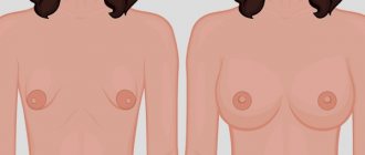
There are 4 degrees of microtia:
Grade I:
A smaller version of the regular ear, with the same physical characteristics and the external auditory canal present.
Grade II:
Partially formed outer ear with a very small or narrow ear canal.
The ear canal may become closed (canal stenosis), resulting in hearing loss. III degree:
Absence of the outer ear of a typical shape (only a small remnant of cartilage and earlobe is present), absence of the external auditory canal and tympanic membrane (auricular atresia).
IV degree:
absence of ear (anotia).
Microtia
What parent doesn't want his child to be born absolutely healthy? Unfortunately, although not often, it happens that the formation of the body does not occur entirely correctly, which can result in various congenital pathologies.
One of these defects is microtia - insufficient development or complete absence of the auricle . This is a rare feature that, according to various estimates, occurs in one in 6-10 thousand people. As a rule, it is unilateral, usually on the right, less often on the left side, but in ~10% of cases bilateral (bilateral) microtia occurs.
The results of scientific research, as well as the practice of plastic and ENT surgeons indicate that in approximately half of the cases the problem is combined with other disturbances in facial proportions, and almost always with atresia (partial or complete absence) of the ear canal and middle ear structures.
↑ Causes of microtia
The etiology of this phenomenon has not yet been studied. Many theories have been put forward, in particular - damage to blood vessels, rubella and taking thalidomide during pregnancy - but none of them has been confirmed. A study of the “genetic” side of the issue showed that a hereditary factor may occur, but in any case is not decisive.
Also, the medical literature specifically emphasizes that microtia is not a consequence of the parents’ poor lifestyle while carrying a child. No matter what “bad” the expectant mother does during this important period - alcohol, caffeine, nicotine, stress, etc. – this cannot be a reason to blame yourself for what happened.
↑ Features of the problem, classification of degrees of microtia
Depending on the condition of the auricle and its individual segments, the following types of microtia are distinguished:
| Degree | Symptoms | Photo |
| I | A slightly smaller auricle with an existing ear canal, which, however, is somewhat narrower than it should be normally | |
| II | Partially underdeveloped auricle with absent or very narrow external auditory canal, accompanied by partial hearing loss | |
| III | The auricle is vestigial, i.e. looks like the rudiment of a normal ear. There is no external auditory canal or eardrum | |
| IV | Complete absence of the ear (anotia) |
With unilateral microtia, the second ear usually works and grows normally, but the child must constantly monitor the development of a healthy organ in order to prevent the possible occurrence of bilateral hearing loss in time. You should also pay special attention to colds in order to avoid ear infections and further deterioration of hearing.
↑ Treatment of microtia, reconstructive surgery
The only, but quite effective, option for solving the aesthetic part of the problem is partial or complete surgical reconstruction of the auricle. This is a complex multi-step procedure that can take up to one and a half years or more. It is performed by a plastic, reconstructive or ENT surgeon.
As a rule, such an operation consists of 4 stages, the sequence and techniques of which may vary slightly:
- Formation of the cartilaginous frame of the future ear. The most suitable material is a fragment of the rib or healthy ear of the patient himself. In addition to your own tissues, it is possible to use other materials: donor cartilage, silicone, polyamide thread, polyacrylic, etc. While there are some advantages of using foreign implants (the ability to recreate the frame before surgery, which reduces the duration of the procedure), there is a high probability of their rejection, donor implants - earlier, artificial - later. Therefore, an implant made from one’s own tissues will be preferable to a foreign one.
- Formation of a subcutaneous pocket in the area of the missing or damaged (underdeveloped) auricle, into which the already formed cartilaginous frame is placed. The engraftment of the frame occurs within 4-6 months.
- From the formed ear block, the base of the auricle is created, and it is given the necessary anatomical position.
- At the last stage, the formed ear block is raised to recreate the auricle, and the tragus is reconstructed using a skin-cartilaginous implant taken from a healthy ear. In the area behind the ear, an unaesthetic defect may form, which is closed by a free skin fold. This stage also takes from 4 to 6 months.
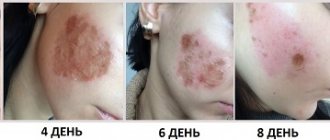
| Photos before and after ear reconstruction for microtia: | |
During the recovery period, a number of complications characteristic of this operation are possible: the occurrence of asymmetry between the healthy and reconstructed ear, reduction of scar tissue and, as a consequence, distortion of the graft, etc. Such problems can be solved by simple repeated surgery. In addition, the reconstructed ear will need to be carefully protected from any traumatic influences throughout its life.
Treatment of microtia involves not only creating an auricle that is visually indistinguishable from the natural one, but also preserving hearing. Therefore, if there is a possibility of functional restoration of the ear canal, then such an operation is performed first, and then the outer ear is also restored. The most difficult case to correct is the complete absence of the auricle - anotia.
↑ Contraindications for surgery
Factors that exclude the possibility of reconstructive surgery for microtia are mainly general surgical:
- the patient’s age is up to 6 years (and, despite the fact that by this age the auricle is already considered fully formed, not all surgeons undertake to operate on patients before adulthood);
- bleeding disorders;
- diabetes;
- exacerbation of chronic diseases;
- cardiovascular diseases.
The last word always remains with the attending physician, who will take into account the general condition of the patient, the degree of manifestation of microtia and possible surgical risks.
Is microtia a genetic disease?
Yes and no.
No:
There are many families who have a child with microtia, but no one on either side of the family has ever had such a child. It appears to be a random event or anomaly occurring during early development. In the case of twins, one child may be born with microtia, but his twin brother will not.
Yes:
Microtia is observed in family generations. Sometimes, an aunt or uncle or cousin has microtia and a new family member is born with the same defect. Relatives may have the right ear affected, and then another family member may be born with a defect in the left ear. Or one family member may have bilateral microtia while the rest have unilateral microtia.
Hearing impairment due to microtia
Because the inner ear develops from a different source than the middle and outer ear, microtia most often does not lead to complete hearing loss. Mostly, patients have a problem with sound transmission. If the diagnosis reveals that the inner ear is normal, the child may be able to use hearing aids to ensure sufficient sound transmission through the blocked area. But if there is no improvement from wearing the device, reconstructive surgery of the middle ear and auricle is indicated. It should be remembered that surgical treatment of such hearing loss is a complex operation associated with a high risk of damage to the facial nerve.
At the same time, unilateral microtia is much more common than bilateral microtia, and a healthy ear provides mono perception of sounds. In this case, correction of hearing impairment is not required, but the appearance of the outer ear will cause discomfort to the patient already in preschool age.
Doctors recommend otoplasty in children and young adults, since at this time the cartilage tissue is most pliable and healing will be easiest. In addition, at the age of 6 the auricle already reaches the size of an adult. Otoplasty before entering school will help the child avoid ridicule from peers.
How will microtia affect my baby?
Microtia affects only external hearing.
Microtia may be accompanied by atresia (absence of the ear canal and often hearing loss), hemifacial microsomia, or other syndromes or genetic diseases such as Goldenhar syndrome or Treacher Collins syndrome. However, microtia does not affect a child's intelligence, motor skills, or success in life. Many parents of children with microtia are shocked at first, but soon realize how lucky they are because there are other children in our world who are born with life-threatening conditions. Microtia affects only the ear itself. Children can have different attitudes towards their ears, some don’t mind such ears, others worry. Try to encourage your child on those days when someone teases him about his ear, and the baby doubts his own confidence and feels upset. Help your child understand that he is no different from other children and can get whatever he wants from life.
Diagnostics
The diagnosis is made immediately after the birth of the child based on examination data.
But not only the statement of this pathological condition is important - it is necessary to make sure that the child does not have other congenital disorders that would develop together with microtia. The mother is asked the following:
- How was the pregnancy overall?
- whether any pathological factors influenced the pregnant woman;
- whether the mother smoked during pregnancy, drank alcohol, or took drugs;
- if there are still children in the family, then whether they, as well as other family members, were diagnosed with microtia;
- whether family members have observed any disorders of the hearing system without visible changes in the outer ear (this may indicate the possible occurrence of pathologies of the middle and inner ear as a result of disturbances in intrauterine development of the fetus).
Particular attention should be paid to the bad habits of the child’s mother during pregnancy, as they could lead to severe hearing impairment, which can only be detected with additional examination.
The physical examination assesses the following:
- during examination - they examine the structures of the child’s facial skull, assess the degree of their development, symmetry, and the presence of possible anatomical disorders;
- during palpation - assess the child’s reaction to palpation in the area of the outer ear and when pressing on the tragus.
Instrumental methods used in examining a patient who is suspected of having a combination of microtia with other congenital pathologies of the hearing organ are aimed at studying the sound-conducting and sound-receiving apparatuses. They are chosen depending on the child’s age and degree of development. Involved:
playing sounds and observing the child’s reaction to them. In this case, speech is addressed to the child or sound objects are used - toys, a clicking clock, a source of music, and so on. Such an examination is carried out on both sides, closing the mouth of the ear canal (if any) on the side opposite to the ear being examined;- otoscopy - the external auditory canal and eardrum are examined using an ear mirror and reflector or using an otoscope (a special device used in ENT practice, which has a built-in optical system and backlight). At the same time, the degree of narrowing or fusion of the external auditory canal and the characteristics of the eardrum are assessed;
- microotoscopy - examination of the same anatomical structures is carried out using a clinical microscope, the study allows you to detect changes that might not have been detected during otoscopy. If there is a congenital violation of the integrity of the eardrum, then it is possible to examine the middle ear cavity;
- computed tomography (CT) - using computer slices, the condition of the structures of the middle ear is assessed, attention is paid to the degree of their development or complete absence - in particular, the auditory ossicles and the temporal bone;
- multislice computed tomography (MSCT) - the method is a more advanced type of CT, with which you can obtain more information and identify possible congenital pathology of the middle and inner ear;
- magnetic resonance imaging (MRI) – used to assess the condition of the auditory nerve;
- radioisotope research - this examination method is used at an older age. The patient is injected intravenously with a special pharmacological drug with attached radioisotopes; they spread through the bloodstream throughout the body, including entering the structures of the middle ear and creating a color image during a tomographic examination. The characteristics of such an image are analyzed and conclusions are drawn about anatomical disorders in the middle and inner ear that have arisen due to disturbances in the intrauterine development of the fetus;
- audiometry – it is carried out on patients starting from preschool age (from 3-4 years). Using special equipment and the examiner’s voice apparatus, sounds are generated and the patient’s threshold for their perception is recorded. Based on these data, a curve is drawn up, with the help of which conclusions are drawn about the state of hearing and assumptions about the congenital nature of the disorders;
- acoustic impendansometry – using special equipment to evaluate the functions of the eardrum and auditory ossicles
Laboratory methods are not decisive in the diagnosis of congenital disorders of the hearing system that accompany microtia, but in some cases they make it possible to assess the condition of the structures of the hearing organ and carry out a differential diagnosis with diseases whose symptoms are similar (confirm or exclude their presence). Usually involved:
- general blood test - an increase in the number of leukocytes (leukocytosis) and ESR indicates inflammatory processes, a sharp increase in ESR, a decrease in the number of red blood cells and hemoglobin - a malignant lesion;
- biochemical blood test - in a number of intrauterine disorders, an imbalance of microelements (sodium, potassium, calcium, magnesium, chlorine and others) is detected, as well as an imbalance in the amount of total protein and the ratio of its fractions.
Surgical options for ear reconstruction for microtia
1. Using your own costal cartilage.
Reconstructive surgery using rib cartilage has been performed since the 1920s. It is a three to four step procedure that involves the use of costal cartilage (rib graft) and a flap of skin to cover the ear (skin graft). Surgery time for each stage can vary from one hour to five hours.
Because a rib ear graft is made from living body tissue, the ear will grow with the person. The reconstructed ear will be slightly stiffer than a normal ear because the costal cartilage tissue is denser, although to the human eye the difference is almost undetectable. The new ear will feel pain, it will bleed when damaged, and it will heal just like the real one.
2. Medpor surgical technique.
In this case, instead of costal cartilage, frames made of porous Medpor polyethylene are used to reconstruct the auricle. This material has been used for craniomaxillofacial operations for more than forty years. Medpor surgery can be performed in one stage, the surgery time varies from eight to twelve hours depending on the complexity of the operation. The Medpor ear is more rigid than the ear with a rib graft. Blood vessels are integrated into the porous openings of the Medpor frame, allowing the reconstructed ear to feel pain, bleed, and heal.
Surgery to restore the auricle and hearing functions for microtia
Microtia II degree Stages of auricle reconstruction
Due to the fact that ear anomalies are usually combined with wired (conductive) hearing loss, and sometimes occur in combination with the sensorineural form, reconstructive operations are a rather complex problem and are planned by a plastic surgeon together with an otolaryngologist. Doctors of these specialties determine the age of the child for surgical intervention, as well as surgical methods, stages and sequence of surgical treatment.
Compared to eliminating a cosmetic defect, restoring hearing in the presence of concomitant atresia is a higher priority. Children's age significantly complicates diagnostic studies. However, if there are visible congenital defects in a child at an early age, the hearing function is first examined using objective methods such as registration of evoked otoacoustic emissions, acoustic impedance measurements, etc.
In children over four years of age, the diagnosis of hearing acuity is determined by the degree of perception of intelligible spoken and whispered speech, as well as through threshold audiometry.
In addition, a computed tomography scan of the temporal bone is performed to detail the existing anatomical disorders.
Determining the age for reconstructive surgery in children also poses a certain difficulty, since tissue growth can change the results obtained in the form of complete closure of the external auditory canal and/or displacement of the auricle.
At the same time, late hearing prosthetics, even with unilateral hearing loss, leads to delayed speech development of the child, difficulties in school learning, psychological and behavioral problems and, in addition, no longer brings significant benefits in terms of hearing restoration. Therefore, surgical intervention is usually planned individually for ages 6 to 11 years. Before surgical restoration of hearing function, especially in cases of bilateral hearing loss, for the purpose of normal speech development, it is recommended to use a hearing aid based on the perception of sound bone vibration, and in the case of the presence of an external auditory canal, a standard hearing aid.
Ear prosthetics
An ear prosthesis is an excellent option that does not require complex surgery. The prosthetic ears look amazing and very real! They can be attached using magnets or glue. You can swim and sleep with dentures. If you decide to reconstruct your ear using an auricle prosthesis made of silicone materials, contact Istok Audio Med LLC
. We cooperate with leading medical institutes and clinics throughout Russia.
119526, Moscow, Vernadsky Ave., 105, bldg. 2. Tkachenko Oksana ext. 525, mobile, Maria Klimacheva ext. 555, mobile,
For a more specific conversation about your case, please send:
- Photo
- Computed Tomography Data
- A copy of the IPR (if there is a disability)
Classification of developmental anomalies of the external ear
Existing classification options for congenital malformations of the auricles are based on the degree of underdevelopment of the external ear.
The Tanzer grading system for underdevelopment of the auricles proposes to classify variants of congenital defects from stage I (complete anotia) to stage IV (protruding ear).
The classification according to the Aguilar system considers the following options for the development of the auricles: Stage I - normal development of the auricles; Stage II – deformation of the ears; Stage III – microtia or anotia.
Weyerd's three-stage classification is the most complete and distinguishes the stages of auricular defect depending on the degree of need for their plastic reconstruction.
Stages of underdevelopment (dysplasia) of the ears according to Weyerd:
- Dysplasia of the first degree
- most of the anatomical structures of the auricle can be recognized. When performing reconstructive operations, there is no need for additional cartilage tissue and skin. Dysplasia of the first degree includes macrotia, protruding ears, and mild and moderate deformities of the ear cup. - Dysplasia II degree
- only certain parts of the auricle are recognizable. Partial reconstruction with plastic surgery requires additional skin and cartilage implants. Grade II dysplasia includes severe ear deformities and microtia (small ear sizes). - Dysplasia III degree
- it is impossible to recognize the structures that make up the normal auricle; an undeveloped ear resembles a wrinkled lump. At this grade, a complete reconstruction using significant skin and cartilage implants is necessary. Variants of grade III dysplasia are microtia and anotia.
What else can you do?
Nothing to do.
You always have the choice to do nothing and accept your microtia ear as it is, to love your ear. In fact, some people with microtia love their small ears and highlight them with piercings and short hair. Some even believe that their small ears are a distinctive feature, something that makes them stand out from the crowd. Visit foreign sites where you will find a lot of useful information about microtia:
In Russia, active communication on microtia issues is conducted on the VKontakte network, the microtia group https://vk.com/microtia
The article was written using materials from the sites: