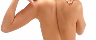
Xerosis , also known as xeroderma , is a symptom whose main symptoms are severe dryness of the skin, its roughness, and sometimes the presence of pityriasis-like scales on the skin.
The main reason for dry skin lies in the disruption of the sebaceous glands (hypofunction). The lack or absence of production of sebum (fat), which is actually the protective layer of the skin from the aggressive external environment, and also which maintains the water balance of the skin, leads to dry skin and its vulnerability to various infections. If there is a lack of sebum in the required amount, the skin not only dries out, it also tightens, peels, and wrinkles.
It has been noticed that if a person has dry and very dry skin, wrinkles appear much earlier, when other signs of aging are not even visible. Of course, not the last factor that leads to premature skin wrinkles is sun rays, which additionally dry out the skin.
Xerosis is dry skin that causes discomfort and unpleasant sensations. Another name is xeroderma. The disease has ICD-10 code L85. Drying out occurs due to a lack of moisture associated with age-related changes, diseases or side effects of medications. Skin xerosis negatively affects a person’s emotional state and creates difficulties in everyday matters. Dryness does not disappear when lubricated with creams and moisturizing oils.
The skin is called the largest and most vital human organ. Its structure includes:
- epidermis;
- dermis;
- subcutaneous tissue;
- adipose subcutaneous tissue.
The unique structure allows you to perform different functions, carry out metabolic, excretory, thermoregulatory vital processes. Defects on the body create cosmetic problems and also indicate the development of diseases in the body.
Causes of xerosis
Dry skin under the age of 25-30 is an inherited problem.
In the case of acquired xerosis of the skin, the reasons may be different and from the photo you can determine how the disease developed.
The problem comes gradually over the years. Contribute to this:
- Unfavorable climate. Excessive cold and heat are one of the main enemies of the skin. In cold weather, the air loses a lot of moisture, and in hot weather, sun radiation destroys collagen and elastin, which are responsible for the elasticity of the skin.
- Appliances. Heaters and air conditioners dry out the epidermis, the top layer of skin.
- Water. When a person comes out of the water, the latter remains on the body and begins to evaporate. As it evaporates, it takes away moisture from the layers of the skin. Therefore, you should not swim, shower or bath frequently.
- Hygiene products. Many of them contain aggressive substances. They destroy the skin's protective barrier - the water-lipid layer. Do not use cosmetics that contain sulfates and alcohol. Also, do not overuse peeling.
- Harmful working conditions. Working in production, for example, at a metallurgical plant, is unfavorable for the body as a whole. The skin reacts first.
- Violation of the regime. Irregular sleep and disordered eating cause premature aging of the skin. Due to dehydration and lack of vitamins. Thus, vitamins A and E are actively involved in the renewal of all layers of the skin.
- Metabolic problems. Promotes hormonal imbalance and improper absorption of nutrients.
- Unhealthy thyroid gland. It affects the activity of the sweat and sebaceous glands, as well as hormonal balance.
- Gastrointestinal diseases. As well as diabetes, hepatitis and cancer of any type.
- Taking glucocorticoid drugs. During a long time.
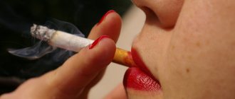
Separately, it is worth mentioning bad habits. Strong tea, coffee, alcohol and cigarettes are not your skin's best friends. They contain many toxic elements that contaminate blood vessels, burden the liver and interfere with the circulation of moisture in the body.
Xeroderma pigmentosum: photos, causes, symptoms and type of inheritance
Xeroderma pigmentosum is a hereditary genetic pathology that impedes the regeneration of skin DNA at the molecular level. The skin becomes too sensitive to ultraviolet solar radiation; there are no enzymes that neutralize the harmful effects.
This is a rare disease, affecting approximately one in 250,000 people. It affects people living in the countries of the Mediterranean coast of Africa and the Middle East. The genetic nature of the disease is due to the fact that a predisposition to pathology occurs when, for religious or other reasons, marriages between close relatives are allowed. A recessive gene appears in the body.
Causes
Of all forms of oncology, 10% are skin cancer. In 90% of cases, it is observed in areas of the body not hidden by clothing. Therefore, the main trigger for skin cancer is ultraviolet radiation.
If the body functions normally, then DNA damaged by radiation should be restored. Recovery occurs in several ways. The first method is that the damaged DNA element is recognized in the chain and removed (cut) using RNA polymerase.
At the same time, a new element is synthesized on the basis of intact template DNA and sewn into place of the removed one.
The second mechanism is the correction of damaged DNA using seven or more enzymes, it is called photoreactivation.
With age, the ability to recover decreases, and the number of damaged DNA elements increases. This is accompanied by changes in the type of oncogenic mutations; genetics stimulates the growth of cancer tumors.
The disease is characterized by a lack of DNA repair.
The following reasons for its occurrence can be identified:
- Autosomal gene.
- Lack of UV endonuclease in body tissues or complete absence.
- Deficiency of RNA polymerase under the influence of ultraviolet rays of a certain length.
- Increased levels of porphyrins and the presence of photosensitivity.
An autosomal recessive type of inheritance is observed. According to studies, genetic correction of xeroderma pigmentosum was presented in the use of retroviral vectors, but the research reached a dead end, since examples of treatment were characterized by oncological neoplasms.
Clinic and symptoms
The first signs appear in early childhood. There are three stages:
- Stage 1: at the age of 2-3 years, flaky spots appear on bare areas of the body from the sun, after which they are replaced by hyperpigmentation and freckles. Further ultraviolet irradiation worsens the symptoms.
- Stage 2: after 2-5 years, signs of skin atrophy, vascular stars and hyperpigmentation of individual areas are observed on the damaged areas of the skin, the skin color becomes mottled. Ulcers appear that are chronically lenticular in nature, and in their place crusts and warts develop. Not only the skin is already suffering, but also the cartilage tissue (nose, ears), connective tissue, which is reticular in nature. Appearance changes, the shape of the mouth, nose, ears changes. The eyes are also affected: blepharitis occurs. The eyelids turn out, the eyelashes become sparse, fall out, stop growing, and the cornea becomes cloudy. The patient suffers from increased lacrimation and photophobia.
- Stage 3: peak of the disease - adolescence and young adulthood. At this stage, various skin neoplasms appear, mostly malignant. Precancerous forms occur: lentigo, limited precancerous melanosis. The growth of warts is characterized by the greatest tendency to degeneration. They have an increased level of oncogenic mutations and metastasis to nearby internal organs.
The neurological form of the disease is distinguished. In addition to skin manifestations, it is distinguished by mental retardation and other signs. These are syndromes named after the researchers: Reed and De Sanctis-Cacchione.
The first syndrome is characterized by slow bone growth, microcephaly, and developmental delay.
Xerodermic idiocy - a synonym for De Sanctis-Cacchione - shows a more severe course and is characterized by the following symptoms:
- reduction of skull bones;
- different degrees of dementia;
- underdevelopment of the brain;
- the appearance of convulsive seizures;
- paresis and paralysis;
- congenital deafness;
- slower growth of bones and limbs;
- underdevelopment of reproductive organs and delayed puberty;
- coordination and reflex disorders;
- inability to bear a child upon reaching childbearing age.
Diagnostics
The main diagnostic method is examination of the skin with a monochromator. This device is widely used to determine the sensitivity of human skin to ultraviolet radiation.
The second method is biopsy - laboratory research of tissue from neoplasms on the skin.
New, typical neoplasms are selected and removed with a scalpel, puncture or using electrosurgical methods.
Small elements are cut off entirely, and large ones are excised wedge-shaped, along with a certain number of healthy cells, thus forming a biopsy specimen. A punctate is taken from a fresh formation with a hollow needle.
Samples are sent for histological examination. Histology makes it possible to draw a conclusion about the need for further treatment, its nature, and allows us to determine the prognosis of the disease.
Histomorphological analysis can reveal the stage of the disease, determine how deeply the tissue is affected, and what is the state of healthy cells.
After the examination, a microscopic description, histological and nosological conclusion is given.
Xeroderma pigmentosum on hands
A differentiated diagnosis will distinguish Xeroderma pigmentosum from radiation dermatitis, although the clinical pictures are similar.
Differential diagnosis is necessary with hereditary and congenital diseases: lentiginosis, congenital dyskeratosis and Thomson's poikiloderma, idiopathic atrophoderma of Pasini-Pierini.
Hereditary lentiginosis is characterized by hyperpigmentation without signs of atrophic changes.
Congenital dyskeratosis affects only men; xeroderma affects equally all genders. Photosensitivity and localization of lesions in open areas does not occur. Manifestations of the disease occur when most sufferers of xeroderma pigmentosum have already died or reached stage 3.
Congenital dyskeratosis is characterized by leukokeratosis of the tongue, cheeks, palate, and aplastic anemia, which is not observed with this disease. Changes in the dermis of a dystrophic nature, detected during histological examination, are not observed in dyskeratosis.
Thomson's congenital poikiloderma is characterized by the distribution of lesions under clothing. Signs of skin atrophy are not so pronounced, cartilage is not affected, and the patient’s appearance suffers to a lesser extent.
Congenital poikiloderma with verrucous hyperkeratosis is characterized by increased sensitivity to the sun. But it is also accompanied by keratoderma and signs characteristic of neurological forms of Xeroderma pigmentosum.
Pasini-Pierini atrophoderma is characterized by various foci, with further atrophic manifestations. There is no possibility of recourse.
Skin lesions have sharp contours and a smooth surface. The color of the rash ranges from brownish to bluish, most often pink or purple. Characterized by a gradual transition to intact skin.
The rash is most often localized under clothing on the back and chest.
The most important clinical sign is hyperpigmentation, possibly a slow chronic course of the disease, slowly progressive in nature.
The disease occurs more often in women, the age of those affected is 20-40 years. Sometimes older people suffer, but more often patients with xeroderma pigmentosum do not live to this age.
Treatment
Clinical guidelines state that the first stage of the disease is treated on an outpatient basis under the supervision of a dermatologist. Mostly synthetic anti-malaria drugs are used. They help reduce the skin's sensitivity to ultraviolet radiation.
Strengthening the patient's immunity occurs with the help of vitamins: PP, A, B.
Corticosteroid ointments are used to eliminate peeling, such as Imiquimod cream and fluorouracil ointment. Warty growths are smeared with cytostatic ointments, they prevent their further growth. 13-sretinoic acid is used internally, it contains isotretinoin.
If necessary, antihistamines are used. Desensitizing substances are administered intravenously: calcium chloride solution 10%, sodium thiosulfate.
The patient lives without the sun, it is necessary to take calcium and vitamin D supplements. Drops with glucocorticosteroids, such as Maxidex, are used for the eyes. Drops such as Artificial tears or Oftagel are constantly dripping.
The condition of patients improves slightly after using reporter enzymes: DNA photolyase, bacteriophage T4 endonuclease locally.
During the period of the most aggressive sun, photoprotective sprays and ointments with the highest filters, lard or quinine ointment are used.
Patients with the neurological form are treated in a highly specialized hospital with the participation of a neurologist and oncologist.
If warts degenerate into malignant neoplasms, the patient is observed by an oncologist. It is good if the patient is simultaneously observed by: a dermatologist, a neurologist, an ophthalmologist, an oncologist, then it will be possible to slightly slow down the degeneration of the growths until they become malignant.
Papillomatous growths of warts have an increased possibility of malignancy, so prompt removal of the growths is necessary. Three treatment methods are used. Cryodestruction – freezing with liquid nitrogen.
Laser therapy is the directed movement of a beam onto an area of skin. It occurs only on the affected areas, clean skin is not affected. Electrocoagulation means burning off warts using an electric current.
The surrounding blood vessels are cauterized, so bleeding does not occur.
Prognosis and prevention
The prognosis is unfavorable. The duration of the disease is 10-20 years, then the young patient dies. A quick diagnosis and compliance with protective measures prolongs life to 40 years, very few live to 50 years, and a few live to 70 years. But still, 75% of children do not live to see the age of 15.
Studying the pathogenesis of the disease provides food for research that identifies cases of predisposition to the disease and promising treatment options. However, there are no drugs for adequate therapy.
You can only alleviate the condition of patients with the help of vitamins and ointments.
Prevention is important to reduce the negative effects of ultraviolet radiation and reduce the risk of tumors that degenerate into malignant ones.
The patient is observed by pediatricians, dermatologists, and later an ophthalmologist joins. A neurologist is involved in the treatment of complex forms of the disease. At the third stage - consultations and observation of an oncologist. Observation by a psychologist is important due to the need to lead a closed lifestyle, associated problems with communication, and appearance as a result of the progression of the disease.
Go outside only in the evening or at night. During the day, wear closed clothing made of thick fabric, a Panama hat or hat, and sunglasses with shields on the sides. For exposed skin areas, special ointments and creams with the highest SPF protection are used.
Cover windows, both at home and in the car, with light-protective films or tinting.
Patients are contraindicated from interacting with carcinogenic factors:
- tobacco;
- radiation;
- mold toxins;
- solvents, petroleum products, asbestos, and other harmful chemicals.
Correct diagnosis of the degeneration of problem areas of the skin into benign and malignant formations is important. Their rapid elimination surgically, using cryodestruction, laser, electrocoagulation and removal of tumors and metastases in internal organs prolong life.
Source: https://onko.guru/termin/pigmentnaya-kseroderma.html
Xerosis. Symptoms
Manifestations of skin xerosis are specific:
- Skin tightness;
- Increased sensitivity;
- Frequent irritation and rashes;
- Redness;
- Invisible pores;
- Coarsening of the skin surface;
- Peeling and cracks (especially in fold areas).
People with xerosis suffer from itching and constant skin tightness. Sometimes there is a burning sensation. The symptoms worsen after contact of the epidermis with water and dry air.
In general, this disease includes four stages of development. Based on their description, you can also determine the presence of xerosis and its development:
- Stage one. The main criterion is functional impairment. The skin loses its protective properties. There is dryness and tightness, which do not cause much discomfort. For now, the problem can be solved with moisturizing creams and that’s enough. Tightness is often felt during facial movements. No wrinkles appear. Sometimes peeling appears after contact with water, sun or dry air.
- Stage two. The appearance and development of hyperkeratosis. Dryness and tightness are always present. Even after the cream. Cause significant discomfort. Small wrinkles begin to appear. Peeling becomes noticeable. Skin sensitivity increases and makes itself felt even with minor influences: contact with warm water, being in a room with an air conditioner running. From time to time, itching and redness bother you. The upper skin layer (epidermis) becomes thinner, its integrity is compromised, and hyperkeratosis (unhealthy epidermal thickening) appears.
- Stage three. Dermal hypotrophy. The peeling is large. Wrinkles are more obvious. Especially on the face (expression wrinkles). The deformation has reached the dermal layer (deeper than the epidermis). Any creams do not work. The skin is inelastic and looks stretched. Hard and rough to the touch. It's cracking. Sensitivity increases several times, inflammation appears.
- Fourth stage. Atrophy of the epidermal and dermal layers. Trophic type ulcers may form on the skin. Signs of premature aging appear.
Read also Natural remedies for eczema
Xeroderma - treatment, symptoms and photos
Xeroderma or xerosis of the skin is one of the varieties of ordinary ichthyosis, which is distinguished by its mild course, positive prognosis and good response to treatment.
It belongs to the group of autosomal dominant genetic diseases, which means it is inherited from the parents. Another name is abortive ichthyosis.
With this disease, a disorder of keratosis is observed, the normal structure of the stratum corneum of the skin is disrupted, as a result of which it peels off and becomes covered with small pityriasis scales.
It is difficult to calculate the exact percentage of patients suffering from this problem, since many people think that flaking is a consequence of excessive dryness of the body, so they do not consult a doctor.
Symptoms of skin xerosis
The main symptoms are dry, rough skin and small scaly scales located on the elbows and knees, and sometimes on the buttocks. These scales are light in color and are easily separated from healthy integuments.
The patient's skin is prone to redness and reacts with increased sensitivity to external influences (itching and burning may occur after applying cosmetics and alcohol, contact with cold air, friction). After water procedures, a feeling of tightness occurs.
The extensor surfaces of the elbows, knees and fingers can become very rough and even crack.
Xeroderma (xerosis of the skin) in children usually goes away after puberty. However, such people must take care of the body throughout their lives, nourish and moisturize it.
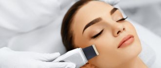
The diagnosis is made by a dermatologist based on the clinical picture and medical history.
Article on the topic: Follicular superficial ichthyosis (keratosis)
Treatment of skin xerosis
Unfortunately, like most diseases of genetic origin, only symptomatic treatment can be used for abortive ichthyosis. It does not eliminate the cause, but helps improve the quality of life (the skin becomes more beautiful, which is very important for female patients).
Oral medications
Aromatic retinoids are recognized as the most effective oral medications. In severe cases, they must be taken for life because symptoms return after stopping therapy.
Because aromatic retinoids can cause significant side effects during treatment, the doctor tries to determine the lowest effective dose to minimize negative effects.
Contraindications for the administration of these agents are liver disorders, excessive concentration of vitamin A in the body and high cholesterol levels.
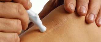
Local preparations
Xerosis of the skin (Xeroderma) requires regular use of exfoliating ointments containing salicylic acid, glycolic acid and urea. They are applied to the affected areas (elbows and knees). The following medications best cope with unpleasant symptoms:
- Keratolite;
- Keratolan;
- Diprosalik;
- Bensalitin;
- Ureotope.
Baths of warm water with the addition of soda or salt (concentration - about 3 percent), as well as collagen baths, are also useful. After the collagen bath procedure, the skin is covered with a thin layer that retains moisture in the epidermis and eliminates dryness and flaking.
At night, it is recommended to make compresses with an aqueous solution of propylene glycol. The product is applied to the elbows and knees, and wrapped with oilcloth on top.
You also need to take care of your body with the help of cosmetic creams, oils and lotions. Apply them every day, preferably after water treatments. Moderate sunbathing, spa treatment, and thalassotherapy bring relief to the patient.
Treatment with folk remedies
You can use all folk remedies that combat excessive dryness of the epidermis. Here are some popular recipes.
- Unsalted lard is mixed with string grass and calendula flowers. Next, all this needs to be boiled in a water bath for 2 hours, strain and cool. You will get a softening ointment.
- Horsetail grass, marigold flowers and chamomile flowers are mixed in equal parts (a glass each) and boiled in a small amount of water. This decoction is added to the bath.
- Sea buckthorn oil with honey and aloe juice is an excellent balm for dry areas of the body. Keep it on for half an hour, then wash off with warm water.
Surely you will find many more natural remedies that relieve unpleasant symptoms.
Prognosis and complications of skin xerosis
Xeroderma does not affect life expectancy, so the prognosis is favorable in all cases. Complications can arise if the patient ignores skin care rules and does not treat peeling areas.
In place of the scales, roughness and cracks appear; they may turn red and itch. The epidermis in this place darkens a little and becomes sensitive to mechanical influences. It's unpleasant, but not fatal.
With appropriate treatment, these complications disappear.
Prevention of skin xerosis
Due to the fact that the disease is genetic in nature, it cannot be prevented. But patients must prevent relapses and not allow their skin condition to worsen.
For this purpose, the techniques that we have already described in this article are used.
Particular attention should be paid to prevention during the change of seasons, nervous stress and any changes in the body (for example, during pregnancy or diet).
Xerosis of the scalp
A particular manifestation of xeroderma is xerosis of the scalp, the causes and treatment of which are determined by the doctor.
In most cases, this is caused by hormonal imbalance. In second place is incorrectly selected skincare products.
Before going to the hospital, you should try to solve the problem yourself:
- Change the shampoo to a softer one - sulfate-free. Or resort to washing your hair with hair balm. The latter should not contain silicones.
- Skip the hairdryer. And other devices and instruments that injure the hair and scalp.
- Drink more water. To replenish the body's lack of moisture.
- Use moisturizing masks for the scalp. Sold in a pharmacy.
And also take into account general advice on the treatment of xerosis.
Xerosis in children
The causes of skin xerosis in children are different, and treatment depends on their nature.
So, the causes of skin xerosis in children may be as follows:
- allergy;
- endocrinological disorders;
- weak immunity.
In most cases, all this is transmitted with genes. To identify the cause, it is worth visiting the appropriate specialists.
It is acceptable to use special children's cosmetics. For example, moisturizing and soothing creams. Sometimes the doctor may prescribe medications.
To combat the scourge on their own, parents can be recommended to use “improvised” measures. This will improve the condition of the child’s skin texture (examples of skin xerosis in children photos).
- start hardening (if there are no contraindications);
- instill a love of hygiene;
- improve nutrition (fresh natural foods, more vegetables, eating at the same time);
- establish a routine (wake up and go to bed at the same time every day);
- ensure healthy sleep (8-9 hours a day, in absolute darkness, in a pre-ventilated room);
- get interested in sports (for example, enroll in some sports section).
Regular spending time outdoors will also benefit your growing body. However, it is better to avoid active sun.
In general, skin xerosis in children is similar to that in adults, regardless of the cause, so the approach to therapy is similar.
Causes
It was previously said that this is a hereditary disease. An autosomal gene is responsible for the development of xeroderma pigmentosum, leading to a congenital defect in excision repair, which is associated with the lack of UV endonuclease activity.
In a cell culture of a healthy person, after irradiation with UV rays at a dose of 10 J/m, up to 90% of thymine dimers disappear from DNA after 20 hours (at a rate of 40,000 dimers per hour), while in cells of patients with XPI, dimers are not removed from DNA at all .
It has not been established reliably who is responsible for the transmission of the autosomal gene - on the maternal or paternal side. But this is always a consequence of closely related ties, starting from the 5th generation.
Treatment of xerosis
When treating skin xerosis, it is worth taking into account all the factors that can cause this disease. Treatment should be combined: medications combined with a correct lifestyle and well-chosen cosmetics. You can also add traditional methods.
Drug therapy is recommended:
- Topical moisturizers (such as glycerin and hyaluronic acid). They retain moisture in the body and attract it from the outside. Promotes better cell division - skin renewal.
- Moisturizing and soothing creams, gels and ointments (usually they contain urea, salicylic and lactic acids). They soften the damaged area of the epidermis and remove dead scales.
- Products containing corticosteroids (indicated only for serious problems and prescribed by a dermatologist);
- Antibiotic medications (in case of infection through the skin).
A little about a healthy lifestyle:
Proper nutrition harmonizes water balance. Therefore, maximum attention should be paid to nutrition. It is worth giving up processed foods - they contain a lot of salt and preservatives.
The latter not only contribute to the rapid loss of moisture from the body, but also disrupt metabolic processes. In addition, there are few natural vitamins.
How to treat?
The disease is treated by a rheumatologist.
biochemical and general blood tests, tests for autoantibodies, cholesterol, bilirubin, creatinine, urea, as well as x-ray and ultrasound examination of internal organs.
In accordance with the results obtained and the severity of the disease, the doctor develops a treatment plan and gives clinical recommendations to his patients.
- It is important to avoid hypothermia and sudden changes in air temperature, winds, direct exposure to sunlight, and contact with household cleaning products.
- you need to give up smoking, drinking alcohol, staying in open water, swimming pools for long periods of time, and visiting a bathhouse or sauna.
- Hot, spicy, salty, and highly chilled foods should be avoided in the diet.
Among the drugs used in treatment, vasodilators, anti-inflammatory, and antisecretory drugs are used. As well as immunosuppressants, enzymes and steroid hormones.
Physiotherapy includes: laser treatment, electrophoresis, ultrasound, ozokerite and paraffin applications, mud wraps, mineral baths.
In the normal course of the disease, people with scleroderma live a long time. But with the progressive development of the disease, 1 out of 8 people die. Therefore, it is very important to start timely treatment and strictly follow all medical recommendations.
Xerosis. Diet
To make dry skin feel better, you should include these products in your menu:
- seafood (contains a lot of polyunsaturated fatty acids);
- vegetables and fruits rich in vitamin C (an antioxidant responsible for skin renewal);
- vegetables and fruits of red and green colors (contain beta-carotene, which is converted into vitamin A. This is a serious antioxidant that does not allow cells to be destroyed);
- clean, fresh water (daily intake for women - from 30 ml per kg of weight, for men - from 40 ml per kg of weight per day).
Traditional recipes:
- Glycerin bath. Restores the epidermal cover. Add 0.5 cups of natural glycerin (non-technical) to a bath of warm water. Take 10-15 minutes.
- Honey-oil bath. A liter of milk is heated without boiling. Add 200 g of natural honey (not store-bought, it is better to purchase it from beekeepers). Mix and gradually add 1 tablespoon of almond oil. The mixture is poured into a bath of warm water. Take - 10-15 minutes. Softens the skin.
- Olive mask. Mix 2 tablespoons each of olive oil and honey. Apply to damp skin after shower or bath. Keep for 20 minutes. Rinse off with warm water. Softens and moisturizes the skin.
How to cure xeroderma
If the diagnosis is known - xeroderma, then you must follow the doctor's recommendations . They depend on the severity of the disease, test results and the general condition of the patient, and should be prescribed on an individual basis.
Primary signs of the disease require periodic examination by a doctor, so outpatient treatment is allowed, with a visit to a medical facility for a follow-up examination. If symptoms of a complication of the disease are suspected or appear, the patient requires hospitalization and appropriate treatment.
In this case, therapeutic treatment may be prescribed - medications that reduce the sensitivity of the skin to ultraviolet rays: "rezoquin", "delagil", or "khingamine". In some cases, sunscreens - ointments, creams - can be useful. Additionally, ointments with corticosteroids may be prescribed for active peeling of the skin or cytostatics for warty growths. Sometimes antihistamines are used - “tavegil”, “diphenhydramine” or “suprastin”.
If malignant tumors are detected, treatment is prescribed by an oncologist. In some cases, surgery may be required. The same method can be used if it is necessary to remove warts.
Not the last place in the treatment of xeroderma should be given to strengthening the human immune system - various vitamin complexes containing vitamins A, PP, B and others can be used.
Prevention of xerosis
To prevent xerosis, a number of conditions should be taken into account:
- Proper sleep is necessary for a person. It restores the body's resources and, accordingly, skin cells.
- Food is of utmost importance. These are the “building blocks” of the body. Skin is formed under the influence of many factors, but its main resource is what we consume. Convenience foods and sweets do not give the skin what it really needs.
- Overheating and hypothermia are harmful. This refers to aggressive environmental factors that cause the skin to “defend itself.”
- Bad habits destroy the body. Starting from smoking to the habit of staying late at the TV screen - all this is stress. And for the skin as well.
Manifestations of skin xerosis, causes and treatment are different (it’s worth looking at photos of different clinical cases). If you approach the problem thoroughly and create a treatment system, dryness can be both prevented and cured.
Causes of skin xerosis
Xerosis can have two main etiologies - congenital (atopic xerosis) or acquired.
If we talk about xerosis, which manifests itself in infants during the first months of life, then it can be a symptom of a mild form of ichthyosis. If we talk about acquired xerosis of the skin, then various unfavorable factors (reasons) can contribute to this.
Read also: What is furunculosis, causes and treatment
Of course, by the cause of xerosis it is fair to designate the cause of hypofunction of the sebaceous glands, which leads to insufficient production of sebum by them, which is manifested by dry skin. Let's look at them.
Causes of xerosis (dry skin):
- Features of the skin – thin skin;
- deficiency of vitamins in the body (hypovitaminosis), especially vitamin A;
- exposure of the skin to direct sunlight (ultraviolet radiation), incl. visiting a solarium, as well as cold, wind, rain, snow, frost;
- frequent bathing in hot water, daily hot showers;
- use of soap with surfactants (surfactants) for washing the body;
- use of household cleaners and detergents without protective equipment (gloves);
- use of alcohol-based cosmetics;
- hormonal changes in the body, age-related changes;
- long-term use of hormonal agents - systemic and external glucocorticoid drugs;
- metabolic disorders;
- violations of the daily routine - work/rest/sleep;
- diseases of the endocrine system, skin, gastrointestinal tract: psoriasis, eczema, ichthyosis, dermatitis, keratosis pilaris, diabetes, gastroduodenitis, hepatitis, liver cirrhosis, hypothyroidism, cancer.
Dry facial skin - causes, prevention and treatment
Dry skin (medical name - xerosis, xeroderma) is a condition of the skin in which dehydration occurs. It manifests itself as a feeling of dryness, tightness of the skin, which often shows signs of irritation, peeling, early wrinkles and signs of aging.
Skin is the largest organ of the human body. Its area ranges from 1.5-2.2 m. Like other organs, the skin has its own important functions: thermoregulatory, metabolic, respiratory, excretory, etc. Dry skin is not only a cosmetic defect, but can also indicate various diseases.
The main reason for dry skin lies in the disruption of the sebaceous glands (hypofunction). The lack or absence of sebum production, which, in fact, is the protective layer of the skin from the aggressive external environment, and also which maintains the water balance of the skin, leads to dry skin and its vulnerability to various external influences.
Symptoms of xerosis
The main symptoms of xerosis (xeroderma) are:
- severe dryness, tightness and irritation of the skin;
- the presence of easily detachable scales on the skin, roughness;
- the appearance of cracks in the skin, especially in places of bends/folds - fingers, elbows, knees;
- coarsening of the upper layer of the epidermis;
- almost invisible pores;
- the presence of wrinkles at an early age;
- a feeling of itching of the skin, sometimes severe, as well as a burning sensation.
Exacerbation of symptoms often occurs after skin contact with cold, water, and soap.
Development of xerosis
The development of xerosis (xeroderma) occurs during 3 stages.
Xerosis 1st degree. Structural changes and wrinkles on the skin are not visible. After skin contact with an unfavorable environment or substances harmful to it, some burning, itching occurs, the skin becomes dry and tightens. Symptoms disappear after treating the skin with a moisturizer.
Xerosis 2 degrees. The structure of the skin is disrupted; in addition to dryness and tightness, peeling, red itchy spots, and wrinkles appear. Symptoms cannot be completely relieved by moisturizing cream alone.
Xerosis 3 degrees. Clear signs of xeroderma appeared on the skin - dryness, tightness, scaly scales, red spots, itching, cracks and wrinkles, especially in the folds. Sometimes swelling appears on the skin. The skin becomes thinner. Frequent skin diseases appear - eczema, dermatitis, infection - staphylococci. Premature aging of the skin occurs. At this stage, serious complex treatment is necessary.
Symptoms of pathology
Sometimes there are five, but more often three stages of the clinical course of the disease - erythematous, or inflammatory, poikilodermic and tumor. However, such a division is very arbitrary and has no practical significance.
The most characteristic signs of the disease are:
- Early age of manifestations of pathology.
- Unusually high degree of sensitivity to sunlight (60%).
- Pigmented rash of red, brown, dark brown and light color, mostly round or oval in shape and with clear outlines (lentigo type). The predominant localization is on the face and neck, less so on the forearms and back of the hands, and even less in areas covered with single-layer clothing.
- Poikiloderma is atrophic areas of the skin in the form of thinning and wrinkling in combination with telangiectasia, alternating zones of decreased and increased pigmentation in the form of a network.
- High risk of developing malignant neoplasms of the skin and internal organs.
- Damage to the visual organs and neurological symptoms.
Cutaneous manifestations of xeroderma pigmentosum
Newborns have normal skin. For the first time, pigmented xeroderma of the face and neck often appears in children in the first month of life. When exposed to sunlight, they develop swelling and erythema (redness). The latter lasts for an unusually long time - up to 2 - 3 weeks, in contrast to a banal sunburn (several days).
Even with short-term and minor irradiation, blisters may appear against the background of erythema, reminiscent of a second-degree burn. Considering the absence of signs of increased photosensitivity (in 40%), such a reaction, as a rule, does not raise suspicions about the presence of xeroderma pigmentosum if there is no medical history.
The first assumptions about this pathology may arise at the age of a child of approximately 1-2 years, and in rare cases of the disease (5%) even after 14 years, when on open skin areas (on the face, neck, limbs) after sun exposure in spring and summer appear excessively pigmented spots resembling freckles (lentigines), with scales of the epidermis.
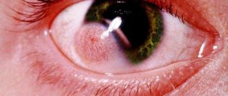
At the same time, increased photosensitivity (photophobia) and eye damage in the form of blepharitis and conjunctivitis often occur, which to a certain extent facilitates diagnosis. Subsequently, eye damage becomes more serious (85%) - the severity of the conjunctival vessels increases, keratitis, corneal opacification, iris damage develops (atrophic processes in the pupillary marginal zone and in the stroma, hyperpigmentation), eversion of the mucous membrane of the eyelids and tumors on them, decrease and complete loss of vision.
In addition, over time, among the spots of hyperpigmentation, areas with reduced pigmentation, telangiectasias, scaly layers, and whitish atrophic scars appear, as a result of which the skin acquires a variegated color. Characteristic symptoms of xeroderma pigmentosum are skin atrophy and hypopigmentation on the dorsum of the nose and the absence of hyperpigmented spots in the chin area.
Treatment of skin xerosis (dry skin)
Treatment of dry skin (xeroderma) includes the following points of therapy:
1. Elimination of the root cause of dry skin; 2. Nourish and moisturize the skin; 3. Local treatment of dry skin areas; 4. Diet; 5. Compliance with preventive measures to prevent dry skin.
Addressing the root cause of dry skin
Treatment of skin xerosis is carried out based on the diagnosis of a dermatologist, who must determine the cause of this pathology.
If the cause of xeroderma lies in diseases of the endocrine, nervous, immune, gastrointestinal tract and other systems, then therapy is aimed at their treatment, and with the right actions, after getting rid of those diseases, the functioning of the sebaceous glands will be restored automatically.
Nourishes and moisturizes the skin. Skin care
Due to the fact that dry skin means there is no protective layer on it, the skin needs to be moisturized and nourished. This must be done to prevent pathogenic microflora, for example, staphylococcus, streptococcus and other bacteria, from entering the body, or the skin layer itself, through micro-traumas of the epidermis. If this is not done, then a secondary infection with its own complications may join the dryness.
Moisturizing and nourishing the skin also gives the skin elasticity, which helps prevent the appearance of cracks, stretch marks, and wrinkles on the skin. Moisturizing also relieves itchy skin.
However, remember that you cannot moisturize your skin with many cheap moisturizers, because... they draw out the remaining moisture from already dry skin. It is best to moisturize the skin with products containing substances such as: vegetable oils (olive, flaxseed, almond), animal fat (bear, greyhound), vitamins, etc.
The best way to moisturize your skin is to drink plenty of liquid – up to 3-4 liters per day. Shouldn't lemonade be the dominant drink?
For your washing, use products without surfactants (surfactants), which disrupt normal sebum secretion, remove the protective film from the skin, thereby increasing dryness of the skin.
Shower gels with a skin moisturizing effect, which contain various oils, incl. ethereal. There is also soap with a moisturizing effect. Yes, these products are usually more expensive, but your health is worth it.
An air humidifier also has a beneficial effect on the skin, especially in winter, when the air in residential areas becomes dry due to heating.
Local treatment of xeroderma
To relieve skin itching, you can take an antihistamine: Suprastin, Claritin, Diprazine.
To relieve itching locally, you can apply a bandage soaked in novocaine to the itchy skin.
For severe itching, you can apply a bandage of weak hormonal ointment - 1% hydrocortisone.
In case of microcracks, in order to prevent infection from entering the body, it is advisable to disinfect the skin by treating it with ointments and creams based on tar and naphthalan.
In case of microbial damage to the skin: if various crusts of a greenish-yellow hue begin to form on the skin, these areas can be treated with antimicrobial lotions: 0.25-0.5% silver nitrate, Brilliantgrun, Rivanol, 0.5 -1% “Resorcinol”, copper sulfate solution. But at the first such symptoms, immediately see a dermatologist!
Read also: Why does my skin itch? Causes, symptoms, treatment, diagnosis
To remove the top layer of scaly epidermis, keratolytic therapy is performed, which is based on treating the skin with creams and ointments with components such as salicylic or lactic acid, urea. These creams soften the skin, remove sebaceous plugs and dead scales, and help normalize the breathing of the epidermis.
Diet for xeroderma
Diet is an integral part of the treatment of many diseases. This is due to the quality of modern food. The fact is that from year to year, less and less healthy food is sold on store shelves. More and more food products are inventions of the chemical industry, artificial products from greenhouses, etc. This food does not contain the necessary vitamins and substances that, when sufficiently supplied to the body, contribute to normal growth, development and human health. Thus, often, diet alone is able to concentrate the body on any failures that have occurred in it, and solve them on its own.
A diet for xeroderma implies the exclusion from the diet of fatty, fried, spicy and smoked foods, fast food, alcoholic beverages, various store-bought chips, crackers, etc.
What can you eat with xeroderma: firstly, drink more fluid, up to 3-4 l/day, secondly, eat foods rich in vitamins and microelements - raw vegetables, fruits, berries, nuts, cereals, fish.
It is worth noting that the least loss of vitamins and other useful substances in food occurs when steaming, so a steamer is an integral household appliance recommended by many nutritionists.
For dry skin, special emphasis should be placed on additional oral intake of vitamins A (retinol) and E (tocopherol). B vitamins are also indicated because most of them, if not all, directly affect the normalization of the cardiovascular system. It’s no secret that low-health food leaves in the human body, especially in blood vessels, remnants of “bad cholesterol”, which over the years disrupts normal blood circulation in many organs, clogging blood vessels, especially disturbances appear in blood vessels with a small lumen (veins, capillaries) . B vitamins help cleanse and quickly remove “bad” cholesterol from blood vessels, blood nourishes all human organs and systems, normal metabolism occurs, and the body begins to function with maximum efficiency.
Additional treatment for dry skin
Beneficial to the surface of the skin, i.e. When treating dry skin (xeroderma), the following procedures are used:
- ozone therapy;
- mesotherapy;
- skin treatment with microcurrents (darsonvalization).
Treatment of scleroderma, drugs
If the patient has focal scleroderma, treatment is prescribed in the form of intramuscular injections of the drug hyaluronidase (an enzyme drug that exhibits affinity for connective tissue fibers). Lidase (another enzyme) can also be injected directly into plaques and spots using ultrasound and electrophoresis.
Additionally, drugs can be prescribed that dilate blood vessels and stimulate microcirculation in tissues: andecamine, nicogipan, kallikrein.
If treatment of scleroderma occurs already at the compaction stage, injections with strong antibiotics of the penicillin group are used. In complex therapy, vitamins A, B15, B and C are also prescribed; they help restore affected skin after stimulation with antibiotics.
Sometimes a dermatologist or infectious disease specialist prescribes hormonal therapy, in which the patient takes thyroidin or estradiol benzoate.
In very rare cases, the patient is prescribed an antimalarial drug: plaquenil or hingamine, which stop the progressive proliferation of connective tissue.
If the patient has systemic scleroderma, intravenous injections of low molecular weight dextrans are used in treatment. The percentage of plasma increases, the blood becomes more fluid and circulates more actively.
The complex prescribes physiotherapy in the form of electrophoresis, ultrasound, diadynamic Bernard currents and paraffin and ozokerite applications. The patient is treated with mud and hydrogen sulfide baths, massage, and therapeutic exercises.
Prevention of xeroderma (dry skin)
- For skin care, use detergents without surfactants;
- Swim in warm water, not hot;
- When using household chemicals, use protective gloves;
- Try to eat more raw vegetables and fruits, foods enriched with vitamins and microelements;
- Avoid alcoholic beverages and smoking;
- Move more;
- Avoid exposure to sunlight in summer, during daytime active sun;
- When sunbathing on the beach, use sunscreen;
- Try to apply less cosmetics to your facial skin; if possible, avoid them altogether, especially if the cosmetics are alcohol-based;
- Don’t let chronic illnesses take their course;
- Avoid stress;
- Wear clothes made from natural fabrics - cotton, silk, but wool can irritate the surface of the skin.
Xeroderma pigmentosum
Considering the statistical data, we can conclude that a disease such as xeroderma pigmentosum occurs in one person out of 250,000. The greatest localization of sick people is concentrated in the countries of the Mediterranean coast of Africa and the Middle East.
Both male and female individuals of different ages are equally susceptible to this disease, but its peculiarity is that most often the primary symptoms appear in pediatric patients. As a rule, the disease in question is inherited and is observed in children whose parents are closely related sexual partners.
Features of the diagnosis of xeroderma and its symptoms
Xeroderma pigmentosum is an autosomal recessive disease, the origins of which are at the genetic level . It is associated with mutation of genes that are involved in the regeneration of previously damaged DNA.
Xeroderma is manifested by sensitivity to ultraviolet radiation and is often the initial stage of cancer.
The first signs of this disease can appear already in childhood - from the second or third year of a child’s life. Initial symptoms may be the following:
- redness;
- peeling;
- inflammation;
- the appearance of freckles;
- excessive pigmentation.
Similar signs are observed on the skin after exposure to the sun. As the time spent under the sun's rays increases, they become more acute.
As the disease progresses, symptoms become more obvious. Among them may be:
- increased pigmentation;
- the appearance of spider veins;
- atrophy of some areas of the skin, which may affect its deeper layers;
- cracks, ulcers, warts appear;
- Possible thinning of the cartilage of the nose and ears, deformation of the mouth, eversion or inflammation of the eyelids - blepharitis.
Such a diagnosis is often accompanied by conjunctivitis, clouding of the cornea of the eye - keratitis, photophobia or photosensitivity.
The advanced stage of xeroderma is accompanied by the appearance of formations on internal organs or tumors - angioma, melanoma or keratoma. Such formations can be either benign or malignant.
There are two forms of complications of xeroderma pigmentosum, which affect the activity of the central nervous system:
- De Sanctis-Cacchione syndrome , which manifests itself in a malfunction of the nervous system. The following symptoms often occur:
- deafness;
- presence of seizures;
- dementia;
- delayed growth and development;
- microcephaly - a decrease in the size of the skull, as well as features of the development of the pituitary gland and cerebellum;
- delayed puberty.
- Reed's syndrome , which often accompanies the diagnosis of xeroderma and manifests itself in children as developmental delays, slowed skeletal growth and microcephaly.
Since the diagnosis of xeroderma pigmentosum is a genetic disease, it is important to know whether the parents or immediate relatives had a similar disease. Such information will be useful when compiling a patient’s medical history when the first signs of the disease are detected.
Causes of the disease
The main reason for such an unpleasant disease is a genetic predisposition transmitted by an autosomal gene from parents to children.
Other reasons are sometimes given as follows:
- Such a diagnosis can be triggered by an increase in the amount of porphyrins.
- Sometimes xeroderma is caused by the absence or deficiency of the enzyme UV endonuclease in the connective tissue.
- An additional reason is a lack of RNA polymerase in the body.
- Another factor may be a sharp decrease in immunity or a slowdown in its formation.
Timely diagnosis of the disease
Timely diagnosis of the disease, accurate diagnosis and competent treatment of such a serious and dangerous disease can provide the opportunity for a successful outcome or improve the patient’s quality of life for a certain period of time.
The primary symptoms of the disease are visible to the naked eye - inflammation, ulceration, peeling of the skin. An additional condition for identifying the disease is that exposure to the sun increases the signs of xeroderma. Such changes in the skin should be a signal for an immediate visit to the doctor.
During a medical examination of the patient, the doctor confirms the presence of the first symptoms of such an unpleasant diagnosis and collects the patient’s medical history - the characteristics of health and the course of the disease, its timing, the history of relatives and the presence of genetic diseases.
Next, laboratory tests are carried out, the results of which make it possible to understand the degree of development of the disease and the stage of tissue damage, which in the future will make it possible to choose the right method of treatment.
Laboratory diagnostic methods include the following:
- The use of a monochromator is a specialized device, the use of which is necessary to assess the sensitivity of the skin to the effects of ultraviolet rays.
- Histological examination or biopsy, which requires the collection of newly formed, atypical tissues, as well as parts of healthy cells - material for research. The procedure can be carried out in several ways:
by electrosurgery method;
If the presence of neoplasms is suspected, the patient is referred to an oncologist to exclude the development of a malignant tumor or to take prompt measures to treat or remove it.
If there are changes in the functioning of the central nervous system, you may need to consult a neurologist.
A comprehensive examination of the patient will be useful - blood and urine tests, examination by an ophthalmologist and dermatologist.
How to cure xeroderma
If the diagnosis is known - xeroderma, then you must follow the doctor's recommendations . They depend on the severity of the disease, test results and the general condition of the patient, and should be prescribed on an individual basis.
Primary signs of the disease require periodic examination by a doctor, so outpatient treatment is allowed, with a visit to a medical facility for a follow-up examination. If symptoms of a complication of the disease are suspected or appear, the patient requires hospitalization and appropriate treatment.
In this case, therapeutic treatment may be prescribed - medications that reduce the sensitivity of the skin to ultraviolet rays: "rezoquin", "delagil", or "khingamine".
In some cases, sunscreens - ointments, creams - can be useful. Additionally, ointments with corticosteroids may be prescribed for active peeling of the skin or cytostatics for warty growths.
Sometimes antihistamines are used - “tavegil”, “diphenhydramine” or “suprastin”.
If malignant tumors are detected, treatment is prescribed by an oncologist. In some cases, surgery may be required. The same method can be used if it is necessary to remove warts.
Not the last place in the treatment of xeroderma should be given to strengthening the human immune system - various vitamin complexes containing vitamins A, PP, B and others can be used.
Patient life prognosis and possible risks
In most cases, patients diagnosed with xeroderma pigmentosum do not live beyond the age of 16-20 years. This trend is associated with the development of severe complications, such as:
- malignant skin melanoma;
- basal cell carcinoma or basal cell carcinoma;
- squamous cell skin cancer;
- septic complications;
- complication of eye diseases – keratitis, blepharitis;
- deformation of external organs - nose, mouth, ears;
- neoplasms of internal organs, both benign and malignant.
Based on statistical data and identified complications, the prognosis of this disease is rather unfavorable, but there is evidence that some patients with this diagnosis lived up to 50 years. The outcome of the disease often depends on the characteristics of a person’s health, the course of the disease and the level of medical care.
In most cases, genetic diseases are accompanied by an unfavorable outcome, which is due to their severity and localization at the gene level. In such cases, modern diagnostic methods and immediate treatment are important, which can slightly improve the patient’s quality of life or cause a favorable outcome.
Source: https://pokozhe.ru/novoobrazovaniya/predrakovye/kseroderma.html
Who is at risk
Elderly people are susceptible to developing excessive dryness when body cells die. The type of dehydration is called senile. Xerosis also develops in those who often come into contact with water or live in cold climates or places with insufficient air humidity.
The disease is classified into two groups:
- Drying with good tone - the epidermis retains smoothness, elasticity and a pleasant color. There are no wrinkles on the surface. But under the influence of irritants, the skin becomes dry and inelastic. This type of dryness is common among young people. This type of epidermis requires constant care.
- Dryness with low tone - the skin is thin, forming early wrinkles around the mouth and eyes. This type of epidermis requires increased care using special cosmetics. Standard methods and creams will not bring visible results.
Having a predisposition to early aging and excessive drying of the body surface, you should carefully care for it using specially developed products. This will help restore elasticity and pleasant sensations.
Stages of melanosis
The disease progresses constantly. The stages are clearly marked by symptoms.
initial stage
For the first time, diagnosis is carried out in children under 2 years of age in the spring and summer. Multiple reddish and pink rashes with easily removable scales, freckles and multi-colored spots appear on exposed areas of the skin.
As the child is exposed to the sun, new rashes appear, but the old ones do not go away. Apart from cosmetic defects, no other problems are observed - no fever, depression, itching, etc.
Xerosis in infants
Dry skin in newborns is not a pathological sign. This is explained by the fact that the baby’s sebaceous epidermal layer does not function for the first time after birth. The discomfort will go away on its own, without the use of additional means, after the body adapts to the changed environment. Also, dehydration in children can be associated with artificial nutrition, early complementary feeding, or a woman’s improper diet during pregnancy and breastfeeding. Increased water hardness and frequent bathing affect the elasticity of the skin.
Dryness of the body often signals problems and diseases of the intestinal tract. Having ruled out possible diseases, it is necessary to combat dehydration of the child’s epidermis. In the first days of life, the skin is very sensitive, easily forms cracks, peeling, redness and itching. This causes discomfort to the baby and interferes with restful sleep.
Dry feet and soles of an infant can cause various infections and bacteria to enter the body and develop inflammatory processes. Insufficiently careful care of a child’s feet leads to the formation of dermatitis and urticaria. Therefore, after each bath, you need to apply a special moisturizer to the baby’s body and massage the legs.
Symptoms of scleroderma, photo
Symptoms differ depending on the form of scleroderma. Some symptoms can behave like separate diseases and can even be treated locally. However, there are several common manifestations of scleroderma for all forms:
- Modification of the skin. The most common lesions are the extremities and face. The skin becomes overly stretched, causing a characteristic shine to appear. There is swelling on the phalanges and between the fingers. Skin changes and swelling occur around the hands and lips. Movement may become painful and difficult.
- Raynaud's syndrome is a lesion of the extremities in response to cold weather or stress. Small vessels become hypersensitive to temperature and impulses from the nervous system, so fingers or even palms go numb due to vasospasm (reduction in the diameter of blood vessels). Against the background of a chronic course, the color of the hands and feet changes, pain in the limbs, tingling and itching may occur.
- GERD is a gastroesophageal reflux disease associated with dysfunction and thickening of the walls of the esophagus and damage to its muscular sphincter. There is damage to the walls due to increased acidity and belching. The peristalsis of the stomach is disrupted, so digestion of food becomes difficult. Problems arise with the absorption of microelements and vitamins through the walls of the gastrointestinal tract. Due to GERD, vitamin deficiencies and minor hormonal imbalances may appear.