- June 22, 2018
- Surgery
- Yulia Lobach
Intervention during surgery is very often associated with complications, including tissue hemorrhages and hematomas. Even without a medical education, it is easy to understand that during this process small blood vessels and capillaries can be damaged. Even the pressure of instruments on the edges of the wound when moving it apart can contribute to this.
Along with the existence of various reasons for the appearance of hematomas after surgery, there are many methods to combat them.
Causes
According to studies, during surgical interventions, eight out of a hundred patients develop hematomas as postoperative complications. Such bruises can appear from subcutaneous hemorrhage immediately after surgery, and sometimes they appear only after several days.
Hematoma after surgery can be caused by the following factors:
- high blood pressure in the period after surgery;
- insufficient blood clotting caused by any diseases or certain medications;
- manifestation of atherosclerosis;
- violation of the integrity of blood vessels;
- vascular diseases or injury;
- non-inflammatory (hemorrhagic) rash;
- the presence of persistent infection;
- very severe diseases of blood vessels and internal organs, such as neoplasms, cirrhosis of the liver and vasculitis (inflammation of blood vessels);
- nutritional problems, lack of vitamins K, B, C and folic acid.
How does a hematoma form?
In most cases, it appears within 24 hours after surgery. There may be several reasons:
- high blood pressure;
- vascular pathologies leading to weakening of their walls;
- problems with blood clotting;
- past traumas;
- use of drugs containing aspirin;
- deficiency of vitamins C, K, B;
- lack of folic acid;
- oncology.
Finally, a hematoma may be found in the patient at a later period, during the recovery process, if he allows himself sudden physical exertion or gets injured.
What danger do bruises pose?
The main risks are as follows:
- first of all, against the background of bruises, infectious tissue lesions can develop;
- often the formed seals do not dissolve for many months and sometimes years;
- scar deformation is observed, which leads to limited mobility.
An internal hematoma that appears in the brain is fraught with damage, often irreversible, and this, in turn, increases the risk of death of the patient.
Types of hematomas and their characteristics
We will consider the treatment of hematoma after surgery later, but for now we will determine their types. A hematoma is a hemorrhage into soft tissue that occurs when blood vessels are damaged.
In the process of cutting tissue, the surgeon can damage the numerous and branched vascular network, which will ultimately lead to hemorrhage. Its main indicators may be: painful sensations, a rapid change in skin tone, the appearance of swelling, and in the presence of internal hematomas, compression is felt, however, all these signs do not always appear and not immediately.
According to location, it is customary to divide hematomas after surgery into four types:
- hematomas located under the skin;
- between muscles;
- hematomas of the chest and abdominal cavity (cavitary);
- located inside the skull.
Types of hematomas
A hematoma itself is an accumulation of blood, both in the tissues and under the skin. Occurs when there is mechanical damage to blood vessels. This often happens due to:
- bruise;
- fracture;
- development of certain pathologies.
Surgery is usually considered the most radical and traumatic treatment option - which is why doctors resort to it in the most extreme cases. Here, when making an incision, the blood contained in the vessels enters the soft tissue, and this is how bruises are formed that appear after operations.
Subcutaneous hematoma
Subcutaneous postoperative hematoma refers to minor hemorrhage into the soft tissues under the skin. Blood may not penetrate the tissue, but rather collect in one place, resulting in the formation of a cavity. In this case, we can talk about a formation in the form of a bag.
Under the skin, a bruise is an oblong or round bruise. There are cases when, in the presence of numerous hematomas, hemorrhages form a cluster of spots.
One indicator is color change. First, red is replaced by purple, and then it becomes yellow-green. Before it disappears, the bruise looks like a brown spot, and remains like that for quite a long time, and leaves no trace behind. If a hematoma forms after an operation of this kind, you just need to wait.
Suture hematoma - treatment
The following remedies will help relieve pain from a suture hematoma:
A warm bath or shower will help treat suture hematoma;
Using cooling or cold compresses. You can apply cold witch hazel, or take a cool shower;
Antiseptic gels and spirea, which include lidocaine, also help with suture hematoma;
Painkillers such as paracetamol, ibuprofen, spasmalgon, no-spa, analgin and the like will help in the treatment of suture hematoma;
You can use lead or zinc ointment with the addition of iodine for suture hematoma;
A compress of a mixture of grape wine and vinegar can also speed up the healing process of a hematoma;
Another good remedy for suture hematoma is brewer's yeast diluted with water and salt. This compress will quickly relieve swelling;
Bodyagi herb can be applied to the site of injury; it will also help the hematoma resolve quickly.
You can simply draw a mesh with iodine on the bruised area three times a day;
A compress made from cabbage leaves and iodine will quickly cure ailments such as suture hematoma;
You can quickly resolve the hematoma using a compress made of dimexide, vodka and water, all diluted in equal quantities, applied to the bruised area and covered with cling film, after which it is fixed with a bandage and left overnight;
You can also make a compress from burdock leaves to treat suture hematoma;
Infusions of medicinal herbs such as chamomile, calendula, millennial, St. John's wort and many others help treat suture hematoma. The main thing is that they all must have anti-edematous properties.
All these remedies are absolutely safe for health, and will only improve and speed up recovery from suture hematoma.
How to speed up the healing of a suture hematoma?
Be sure to keep the seams completely clean so as not to cause an infection. And, of course, do not wait until the hematoma goes away on its own, but engage in treatment. It is best to consult a doctor before treatment. If the hematoma does not go away for a long time, or its condition worsens, a visit to the doctor is required!
- This is a cavity formed as a result of injury and filled with blood or clots. Occurs when blood spills into tissue from a damaged vessel. It is a dense or fluctuating tumor-like formation, painful on palpation, accompanied by swelling and discoloration of the skin. With a deep location, it manifests itself as a violation of the shape and a local increase in the volume of the affected area. Pathology is diagnosed based on complaints, anamnesis and external examination. In doubtful cases, ultrasonography is prescribed. Treatment is local conservative measures; in severe cases, opening and drainage are indicated.
Internal hematoma
This type of hematoma is an extremely dangerous form of hemorrhage, since in addition to damage to organs, large quantities of blood fill cavities or can accumulate between tissues.
In humans, these symptoms begin to appear with a sharp drop in blood pressure, the skin becomes pale, and pain appears at the site of the operation. Blood flows out through the drains in huge quantities. The condition is so dangerous that if the person is not put on the operating table at the right time and the bleeding vessel is not sutured, then everything can end sadly. For example, a hematoma after hernia surgery may be of this type.
Treatment
Treatment measures will also directly depend on the depth and volume of the hematoma, as well as the presence or absence of its complications. Simple superficial hematomas, as a rule, do not require treatment and go away on their own, completely resolving in 14-20 days. The patient does not have to stay in a medical facility for this period. He can be at home and follow simple recommendations, which we will give below.
Intense hematomas must be subject to surgical treatment - the cavity is opened, emptied and drained, and the vessel feeding it is ligated or coagulated. For bruises in internal organs, special hemostatic sponges and adhesives are widely used. In complex therapy, various hemostatic agents are used, for example, Etamzilate, Aminocaproic acid, Tranexam.
Infected and festering hematomas are also subject to surgical treatment. Infected tissues and pus are removed from the cavity, the tissues are treated with antiseptics, and, if necessary, a rubber tube or strip is left in the hematoma cavity - a drainage through which the pus will flow out. The patient must be prescribed a course of antibiotics to suppress pathogenic microorganisms.
And finally, some tips. Cold on the area of the postoperative wound. This method is effective in the first hours after the formation of a hematoma. Cold promotes spasm and closure of blood vessels and prevents bleeding. The use of absorbable agents - compresses with heparin ointment, bodyaga. There is evidence of the effective use of leeches - hirudotherapy.
Physiotherapy is very effective for rehabilitation in the postoperative period: magnetic therapy, ultrasound, electrophoresis with lidase, darsonvalization.
Dosed physical activity, quitting smoking, alcohol and coffee and controlling blood pressure. Share:
Brain hematoma
Bleeding inside the skull can occur after injury or surgery. This phenomenon is deadly, as there is a possibility of irreversible damage to human brain tissue. Therefore, it is imperative to immediately diagnose and treat these cranial hematomas. The following types are distinguished:
- subdural, associated with traumatic brain injury;
- epidural;
- hematoma inside the brain.
The first hard shell is located between the brain and the bones of the skull. At the end of the operation, accumulated blood is found between this membrane and the skull. In medicine, this is called a subdural hematoma. Such conditions lead to increased pressure on the brain and changes in its functioning for the worse. There are cases when this type leads to death.
Possible complications
Even a small and seemingly harmless bruise, if treated incorrectly, can lead to serious consequences, for example, suppuration of the bruise. Internal hematomas are sometimes accompanied by infection and purulent inflammation. When damaged skin peels off at the site of a bruise, traumatic lumps (cysts) can form, which can only be removed through surgery. If bed rest is violated after surgical treatment, repeated accumulation of blood in the joint (hemarthrosis) is possible. Severe injuries and extensive bruising can lead to tissue necrosis.
Under no circumstances should hematomas, especially large ones, be left without the attention of doctors.
Of course, accidental injury cannot be prevented in advance, but its consequences can be minimized. To strengthen blood vessels, muscles and bones, you need to eat a balanced diet, lead a healthy lifestyle and exercise - all this together will help prevent the development of serious complications after bruises.
What to do if you didn’t hit yourself anywhere, but bruises appear on your skin?
Many have heard about such a complication after medical procedures and operations as a hematoma. In our article today we will talk about what a hematoma is after surgery, why it forms and how to deal with it.
Hematoma is literally translated from ancient Greek as “tumor from the blood.” A hematoma or, in common parlance, a bruise is an accumulation of blood in a limited cavity. It is important to understand that a cavity filled with blood is not natural - that is, blood, pouring out of damaged vessels into the surrounding tissues, itself forms this space. The accumulation of blood in natural cavities: the cardiac sac, pulmonary pleura, sinuses, uterine cavity, and so on is a completely different situation.
Hematomas are divided into several types depending on the method of formation. c - that is, received from blows, most often with hard blunt objects on soft tissues. When struck, blood vessels of various sizes and calibers are damaged, blood begins to pour out of the damaged vessels, pushing apart the soft tissue under pressure - an accumulation of blood is formed.
Postoperative is a special type of traumatic hematoma. Hematomas after surgery are, in principle, a common postoperative complication or even a variant of the normal course of the postoperative period. With any surgical intervention, one way or another, the vessels of the skin, subcutaneous fat, muscles and other soft tissues are damaged. Of course, at the end of the operation, the doctor restores the integrity of the tissue as much as possible and sutures or cauterizes large vessels. However, due to the peculiarities of the operation or postoperative period, as well as the individual characteristics of the patient’s body, sometimes after surgery blood may leak slightly through the stitches and form hematomas.
Spontaneous is a special type of formation, characteristic of people with vascular diseases, hereditary or acquired diseases of the blood coagulation system. Various vasculitis - inflammatory diseases of blood vessels, abnormalities in the structure of the vascular wall and even banal atherosclerosis lead to increased fragility of blood vessels. Such affected blood vessels are prone to spontaneous rupture and the formation of hematomas with complete rest of the patient or minimal impact on the tissue - touch or compression. With a low level of platelets - thrombocytopenia, hereditary blood diseases, hemophilia and others, spontaneous tissue hemorrhages can also occur, caused by minimal damage or even against the background of complete well-being.
Subcutaneous hematoma of the thigh
Depending on the location of hematomas in the body, there are several localizations. Subcutaneous hematomas or those same “bruises” are the accumulation of blood in the subcutaneous tissue. This is the simplest type of hematoma, however, with a large area and depth, such formations can also be dangerous to the patient’s health.
Muscle hematomas are an accumulation of blood in the muscle under the fascial sheath. Such accumulations of blood are often combined with subcutaneous bruising.
Subserous hematomas are deep types - these are accumulations of blood in the soft parenchymal organs of the abdominal cavity and retroperitoneal space under their serous capsule - in the liver, spleen, kidney, uterus, etc.
Intracranial - these are accumulations of blood in the thickness of the brain substance. This is the most dangerous type of hematoma, causing severe consequences for the patient. Such hematomas present real difficulties for doctors in terms of treatment, diagnosis and rehabilitation.
Speaking about characteristics, we need to remember the following distribution. Tense or throbbing. This is the most complex form of hematoma, since blood continues to pour into their cavity, pushing the tissue further apart. Such pulsating hematomas, increasing in size, can compress neighboring structures and cause severe pain. Such formations require urgent surgical care - opening, drainage and suturing or coagulation of the feeding vessel.
Unstressed or stationary. Hematomas, the supply vessel of which has closed, no longer increase in size, the blood in their cavity gradually coagulates, stratifies into fractions and gradually resolves - the process of so-called “hematoma blooming” occurs. This is a rather interesting evolutionary process. It can be clearly observed in the example of subcutaneous hemorrhages. At first, the bruise is purple-cyanotic, after 6-7 days it becomes greenish, then, after another 7-9 days it becomes yellowish. These color changes are caused by gradual transformations of the red blood pigment - hemoglobin, and are widely used in forensic medicine for examination and determining the age of injuries.
Based on the presence of complications, two types are distinguished. Complicated are accumulations of blood that can undergo secondary changes: the addition of bacterial flora, infection and suppuration. Complicated hemorrhages also include intense types. Uncomplicated - that is, their cavity is stationary and goes through the usual stages of resorption.
Treatment of hematoma after surgery
To treat the listed pathologies, a certain approach is required, and repeated surgery is not excluded. Treating these types of hematomas yourself is extremely dangerous. Under such conditions, the blood can quickly pick up an infection that penetrates from the surgical wound. A large hemorrhage can lead to extensive blood loss. Decomposition products can cause intoxication of the body.
The general danger is that:
- there is a risk of infection;
- a compaction appears at the site of the hematoma and does not disappear over time;
- the shape of the tissue changes, and the scars that appear in places of accumulation negatively affect their mobility.
When using modern laser surgery techniques, as well as the use of special instruments that can cauterize blood vessels in the wound during surgery, the risk of infection is reduced. We will talk further about in what cases anti-inflammatory ointments and other means of therapy are used.
After surgery, minor bruises will resolve on their own.
Applying cold helps stop bleeding as the blood vessels constrict. Applying a pressure bandage helps localize the leak on the arm or leg. Using a puncture and a syringe, the entire contents of the hemtoma are removed.
These methods are effective for superficial blood collection. A small incision, the so-called drainage, allows you to remove blood clots if the hematoma has coagulated or taken the shape of a bag.
Heparin-containing gels and anti-inflammatory ointments are excellent for treating skin with a small hematoma after surgery. They are applied to the bruise several times a day until the effect is completely gone. Physiotherapeutic procedures complement the treatment and help eliminate compaction and swelling.
For repeated prevention of hematomas, compression hosiery is used. A blood clot should form. And to achieve this effect, it is necessary to immobilize the limb. If bleeding is severe, the doctor will prescribe medications that increase blood clotting.
How to remove a hematoma
For the most part, minor bruises resulting from surgery will go away on their own. To speed up the process, ice is applied to the sore spot.
When a hematoma appears on the leg, a pressure bandage is used. Here, bruises often occur if a phlebectomy or removal of veins damaged by varicose veins was performed.
Small subcutaneous bruises are treated with simple folk methods. In particular, compresses are used:
- from vodka pure or with the addition of vinegar;
- saline solution;
- mustard mixed with radish juice.
Drug therapy involves the use of drugs:
- stopping bleeding;
- antibacterial (for prophylactic purposes).
Any ointment containing non-steroidal components will also work. External treatment allows:
- relieve swelling;
- reduce tissue reactivity;
- alleviate pain syndrome.
It is also advisable to prescribe physical therapy to such patients. They must wear compression garments.
Sometimes puncture helps remove the hematoma. In this case, a needle is inserted into the localization site and thus the accumulated liquid is removed.
In severe situations, when conservative methods do not bear fruit, an operation is performed during which the hematoma itself is removed, and the vessel that has not closed is also cauterized. Upon completion, drainage must be installed for at least 3 days.
Measures to reduce the risk of hematoma formation after surgery
So, to avoid a hematoma after surgery, what should you do? For this purpose the following is carried out:
- Examination of the patient for the presence of blood-related diseases, in particular those related to blood clotting.
- Elimination of drugs that cause a decrease in blood clotting.
- Operations must be carried out with the obligatory restoration of blood loss.
- Damaged vessels must be carefully coagulated.
- After completion of the intervention, before suturing the wounds, it is necessary to check the cavity of the coagulated vessels.
- During the recovery stage, it is necessary to strictly follow the doctor’s requirements, and this must also be done at the postoperative stage.
Do not forget that a hematoma is not so harmless and can negate the result of surgical actions. It is necessary to listen to doctors to avoid complications that may prolong the recovery period.
Postoperative
Postoperative hematomas
Postoperative hematomas are a completely special type of hematoma. They can be of absolutely any type from the above classification: subcutaneous, cavitary, tense, and infected.
It is important to understand that most often such “bruises” are a normal stage of the postoperative period. This is not the fault of the surgeon, nor the suture material, nor postoperative care - these are only the individual characteristics of the patient himself, the nuances of the operation itself and the course of the postoperative period.
Of course, there are factors that predispose to the development of bruises after surgery and medical procedures.
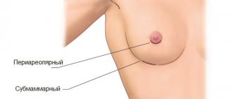
Features of operations. Operations performed on areas of the body with a pronounced vascular network and richly supplied blood organs - face, mammary glands, thyroid gland, as well as on the vessels themselves - for example, leg veins, pelvic vessels. That is, for example, a hematoma on the leg after surgery for varicose veins is a completely normal occurrence.
The patient is taking special medications that affect the coagulation system: Warfarin, low molecular weight heparins, antiplatelet agents. It is recommended to discontinue such drugs at least a month before the proposed operation, but this is not always possible due to the patient’s health.
Arterial hypertension. This disease in a patient causes excess blood pressure in the vessels to push blood into the surrounding tissue, forming a bruise. Individual characteristics of the vascular wall and coagulation system, including the diseases listed above: thrombocytopenia, vasculitis and others.
Smoking. Smokers often have increased fragility of blood vessels and blood clotting characteristics, so the formation of bruises and post-operative bruises is much more common.
Drinking alcohol. Alcohol promotes vasospasm and increased blood pressure, which predisposes blood to leak from vessels that have not yet recovered into the surrounding tissue. Failure of the patient to comply with the doctor's recommendations. Early physical activity, improper dressings, and refusal to take medications can contribute to the formation of bruises and other postoperative complications.
Therapy methods
Removing a hematoma using conservative methods takes a lot of time. The body needs a significant amount of energy and resources to break down its own blood at the site of hemorrhage. Also read -.
Subcutaneous hematomas without complications should be treated immediately. The first thing to do is apply cold immediately after the injury. Thanks to the cold, the vessel narrows, bleeding stops, and the area of the bruise does not increase. The cold should be kept for about 20 minutes, after which ten-minute breaks should be taken. You can apply a pressure bandage in the area of manifestation.
Non-steroidal anti-inflammatory drugs are used to relieve pain. You should not take Aspirin because it thins the blood.
After a hematoma has formed from the blow, special heparin ointments or bodyagu ointment can be applied for treatment. Such products will speed up the resorption process. Indications for surgical removal of hematoma:
- big sizes;
- severe compression of tissues and muscles;
- suppuration;
- hematomas of internal organs that do not respond to conservative treatment.
The operation is performed immediately if the hematoma threatens the patient’s life, for example, there is compression of the brain or its parts, or compression of large arteries.
You cannot open bruises yourself, as this can lead to infection and continued bleeding.
Traditional methods
Each of us knows several folk ways to get rid of a hematoma. If the hematoma does not resolve for a long time, you can speed up this process with the help of traditional medicine.
Treatment at home can be carried out using a leaf of white cabbage. You need to take a sheet, make cuts and apply it to the bruise overnight. The notches are needed so that the juice comes out better.
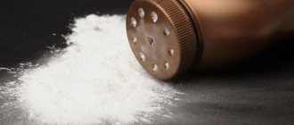
To remove an unsightly blue stain, you can use salt lotions. To do this, you need to melt 150 grams of salt in hot water and soak a bandage in it, then apply a bandage for 12 hours.
Onion mixture works great. Chop the onion and add 3 tablespoons of salt to it, stir. Apply 2 times a day to the bruise. Treatment period is up to 5 days.
A mixture of honey and aloe in a 1:1 ratio is very popular. Apply 2 times a day.
Grated plantain leaf has a good effect. Apply the resulting pulp to the site of the hematoma. Also use castor oil lotions.
All folk remedies are aimed at improving blood flow and accelerating metabolic processes, which stimulates faster resorption of blood clots.
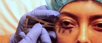
How long do bruises last after blepharoplasty?
Hematomas become most severe during the first two days after surgery. After this period, the bruises gradually decrease. The amount and duration of recovery for each patient is determined individually. Sometimes bruises are localized on one side of the face - left or right. This is absolutely normal, although it happens less often.
The weakest bruises last two to three days. The most severe hematomas can persist for up to two weeks. Such periods are completely normal, and there is no need to panic and wonder what to do with bruises after blepharoplasty. Simply listen to your doctor and carefully follow all instructions he gives you to ensure a great aesthetic result.
Symptoms
One of the earliest and longest lasting symptoms is pain. Pain occurs after swelling and may be pulsating or pulling. The intensity of the pain depends on which nerve endings have been compressed.
During injury, many cells are destroyed, in other words, alteration occurs. When a cell dies, many biologically active substances (BAS) are released, which irritate the receptors and cause a burning sensation and itching. Patients complain of decreased function of nearby muscles and the appearance of limited edema. Due to the release of biologically active substances into the blood, body temperature rises to subfebrile levels. An old hematoma, when disintegrating, can also raise body temperature.
If the swelling does not disappear for a long time, and the bruise does not change color, you need to consult a doctor. This can occur when bacteria multiply in the cavity formed by the hematoma. If a hematoma appears after a blow to the head, in addition to pain, as a rule, symptoms of damage to the central nervous system are observed: nausea, vomiting, loss of performance. It is necessary to examine the patient for the presence of a concussion or other injuries to the brain or skull.
Also, with internal bruises, dizziness, loss of consciousness, decreased blood pressure, and twitching of small and large muscle groups may occur.
If a child develops a bruise two to three hours after being hit, hemophilia should be suspected and tested for clotting factors.
Symptoms
Hematoma bloom
Clinical manifestations depend on the location: subcutaneous or deep. For the subcutaneous, the clinic is obvious:
- Change in skin color depending on the time of formation: from bluish to yellowish.
- Swelling. Increasing edema and swelling in a tense hematoma indicate its suppuration.
- Pain at rest, aggravated by pressure. The progression of pain also indicates a tense or pulsating hematoma, as well as infection and suppuration.
- Temperature increase. A local increase in skin temperature at the site of the hematoma can be observed with normal superficial effusions. General heat and fever are characteristic of infection and suppuration of hematomas, especially large ones.
For deep hematomas of muscles and internal organs, the clinical picture is more hidden:
- The patient is bothered by pain in the projection of the surgical wound, which intensifies with pressure and movement.
- An increase in temperature is also typical for infected hematomas.
- Deep pulsating hematomas, in addition to increasing pain, are characterized by symptoms of ongoing bleeding: weakness, dizziness, pallor, dry mouth, low blood pressure and high pulse.
- Sometimes, with extensive bruises after operations on internal organs, symptoms of compression of adjacent organs by the cavity with blood may be observed. For example, after surgery on the uterus, a mass can put pressure on the bladder, impairing urination.