Umbilical hernia in adults - reviews of the operation, symptoms and treatment
Methods of performing surgery to remove an umbilical hernia: laparoscopic, open, hernioplasty, laser removal. ⭐️⭐️ ⭐️ ⭐️ ⭐️ Duration of surgery, rehabilitation and possible complications. Forecasts and patient reviews
Read advice from our experts
Symptoms of an umbilical hernia are:
- compaction or protrusion in the navel area;
- expansion of the umbilical ring;
- pain in the middle of the abdomen during physical activity, coughing;
- nausea.
The disease can occur at any age in both men and women. But more often, umbilical hernia occurs in infants and people over 40 years of age.
Weakness of connective tissue and abdominal muscles predisposes to the appearance of a hernia in adults. Protrusion can occur with increased physical activity, heavy lifting, or chronic diseases accompanied by coughing. Excess weight, constipation, pregnancy, and ascites (accumulation of fluid in the abdominal cavity) also contribute to the formation of a hernia.
An umbilical hernia in infants can be congenital (due to improper formation of organs in the womb) or acquired. The latter occurs with frequent increases in pressure inside the peritoneum due to crying and slow healing of the umbilical ring. In some cases, infants' hernias are repaired non-surgically - with the help of massage.
The primary examination of a hernia consists of a visual examination of the patient by a general practitioner. Inspection and palpation are carried out in a horizontal and vertical position. If a hernia is detected, the doctor prescribes further examinations:
- Ultrasound of the abdominal organs;
- herniography – diagnosis using a contrast agent injected into the peritoneum;
- FGDS (for the study of a complex of diseases of the gastrointestinal tract);
- computed tomography.
To clarify the shape and size of the hernia, ultrasound diagnostics is sufficient. However, for a more detailed study and identification of concomitant diseases, additional methods are used.
There are a number of contraindications to surgery for umbilical hernia in adults and children. Therefore, it is necessary to carry out examinations in advance, the results of which may force the doctor to reschedule or cancel the intervention. Preparation before surgery to remove an umbilical hernia includes the following tests:
- ECG;
- general blood analysis;
- Analysis of urine;
- coagulogram;
- fluorography;
- tests for HIV, hepatitis, syphilis.
Contraindications to surgery to remove an umbilical hernia are:
- severe cardiovascular disease, recent heart attack or stroke;
- pregnancy, especially 2-3 trimester;
- old age – from 80 years and older;
- cirrhosis of the liver;
- renal failure;
- diabetes;
- varicose veins of the esophagus;
- infectious diseases in the acute stage;
- blood clotting disorder.
Hernioplasty is considered a simple operation, especially for small hernias. But even in emergency cases, when the hernial sac is strangulated or grows, surgical intervention, as a rule, goes well and is successful for the patient.
Removal of an umbilical hernia in adults and children can be performed using the traditional method (using tissue tension, without the use of an implant) or with the installation of an endoprosthesis . The implant is a mesh that strengthens the weakened part of the peritoneum and promotes its overgrowth with new tissue. Endoprostheses are made from different materials. Some of them dissolve over time, leaving behind their own tissues that have grown on top. Others remain in place and can be removed if necessary. Surgery using an endoprosthesis helps reduce the risk of relapse by strengthening the abdominal wall.
Laparoscopic method
About 25 years ago, it became possible to use a gentle method for removing an umbilical hernia - laparoscopic. Now this is the main and safest way to treat hernias. The operation is performed through small incisions, into one of which a camera is placed, and into the other - surgical instruments. Due to its low invasiveness, the risk of adhesions is reduced and the postoperative period is shortened. Sutures after laparoscopy are practically invisible. Laparoscopic surgery for umbilical hernia in women is performed during the intermenstrual period. With laparoscopy, both the tension method of closing the hernial orifice and the installation of a mesh implant are possible.
Single-port laparoscopy or the single-incision SILS method has been used relatively recently and has quickly gained the sympathy of both surgeons and patients. The disadvantage of this method is that the puncture hole is much larger than the standard ones.
Hernioplasty using Sapezhko, Lexer or Mayo methods
These are traditional methods of hernioplasty, which involve closing the hernial orifice using tissue tension. There are some differences in the course of operations and indications for them.
The Mayo method is more often used in overweight people. Two crescent-shaped incisions are made around the navel to capture excess fat deposits. Then the hernial sac is cut, adhesions and necrotic tissue are removed, and the internal organs are set into the peritoneum. The edges of the hernial sac are sutured horizontally. The aponeurosis (the so-called plate of connective tissue that gives strength to the abdominal wall) is sutured.
During surgery using the Sapezhko method, incisions are made in the form of vertical lines. After placing the organs in the abdominal cavity, layer-by-layer stitching of tissue is performed. Otherwise, the procedure is almost the same as the Mayo method.
The Lexer method is suitable for cases where the navel and hernial sac are inseparable. If necessary (large size of the hernia), the navel is removed during the operation. The doctor makes a crescent-shaped incision around the tumor. When the hernial sac fuses with the navel, it can be difficult to release its contents. The bottom or neck of the hernial sac is opened, and the organs are placed in the abdominal cavity. A suture is placed on the neck, and the sac itself is cut off. The hernial orifice is closed using the Lexer method by applying a silk suture to the aponeurosis around the ring and several sutures to the rectus abdominis muscles. After this, the skin flap cut out at the beginning of the operation is returned to its place and secured with interrupted sutures.
Types of umbilical hernia repair
There are types of plastic surgery, which are selected at an appointment with a surgeon in agreement with the patient.
1. Intraperitoneal method. It is used for embryonic disorders in the first month after the birth of a child. Opening the hernial sac, returning organs to physiological places. Excision of the sac. Stitching.
2. Mayo method. Suitable for children over 6 years old and adults. Incision of tissue near the umbilical hernia, its detachment. Opening of the hernial sac. Installation of organs in physiological places. Ligation of the neck, removal of the sac. Stitching. Postoperative recovery takes a long time.
3. Use of mesh implants. The course of plastic surgery is similar to the Mayo method. The difference is the application of a mesh to the area of organ prolapse to eliminate the risk of relapse. Fixation of the mesh using non-absorbable sutures or a surgical stapler. The mesh is made from hypoallergenic materials that are not rejected by the body and do not decompose.
4. Laparoscopy. The surgeon makes punctures, inserts tubes and an endoscope with a camera to examine the surgical field. Through the second hole, tissues are captured, the surgeon opens the tissues, and puts the organs in place. An implant (mesh) is inserted and installed. Sutures are placed through the third hole using a stapler. The advantage of the method is the rapid postoperative recovery period.
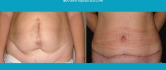
Abdominoplasty, removal of umbilical hernia with navel transfer
before and after photos, plastic surgeon unknown
- Operation:
Abdominoplasty - Details:
Abdominoplasty, removal of umbilical hernia with navel transfer - Result after:
1 year and 9 months - Surgeon:
Surgeon unknown
I had spinal anesthesia, by the end of the OP I began to hear everything, how they were admiring my belly button! I woke up normally, but was half asleep! And then I slept some more, and then... I even ate!
After 2.5 months
2.5 months after the OP, I can finally show the result.
And they cut off a flap of my skin and the blood flow had not yet returned to normal. It’s already 3 months after the op, but the numbness in the suture area still remains.
After 9 months
Six months later
The stitches stopped bothering me after a few months, I started losing weight immediately after the surgery, because... the abdominal muscles were sewn together, when I ate food in the usual amount, I began to burst 30-40 minutes after eating. For a long time I couldn’t understand why? But then I realized that now the stomach had little space in the abdominal cavity and began to eat in small portions. The weight began to decrease ,I lost 4.5 kg in about 1.5 months!
Of course, you need to rest! In my case, it was 2 days and I had to do something myself. Fortunately, my parents and I live now, so my mother didn’t work and took all the responsibilities on herself! But you still have to pace yourself, because it’s also hard to lie down all the time, everything becomes numb. And you get tired quickly, your back starts to hurt - you had to walk in a half-bent state. I had to wash my hair over the bathtub only with someone’s help. And then lie down and rest! And in general, when you do something, time passes faster and the muscles become toned. You can’t stay put for long!
It became active after about 10 days. But this concept is still flexible. I straightened up and began to walk normally, did everything around the house, but fatigue was still present, only after a month I began to forget about the OP
You will have to walk in a bandage for 2-3 months. This is not even discussed.
After 1 and 9 months
And here is the bottom seam. but you are unlikely to see him
It was hard for two weeks, I wanted to lie down all the time. the swelling persisted for a long time.
1 photo seam under the underwear; 2 photos - the seam itself (and more than one) after 2.5 years. Let me remind you: I had an abdomino with two seams - vertical and horizontal
Protruding and distended navel after childbirth
In the last weeks of pregnancy, the navel often protrudes beyond its “hole”. The growing uterus puts pressure on the muscles of the anterior abdominal wall. The skin stretches under the influence of relaxing pregnancy hormones. As a result, after giving birth, a woman discovers that her navel is sticking out. A “hood” of skin hangs over it.
It takes an average of 6-9 months to restore your figure. Over time, both muscles and skin return to normal. But a slightly bulging belly button and overhanging skin may be permanent. This depends on many factors: predisposition to stretch marks, the strength of the abdominal muscles, and the quality of nutrition.
Umbilical hernia in adults
An umbilical hernia is a pathological condition in which abdominal organs protrude through the umbilical ring. Umbilical hernia in adults is not a common occurrence (about 5-12%) - it is more typical for newborns and infants. The main cause of this pathology is weakness of the anterior abdominal wall, which can develop due to various factors.
In this article, we will look at the main symptoms and main reasons why an umbilical hernia occurs in adults, what treatment methods exist, how the operation is performed, and also show what an umbilical hernia looks like in the photo.
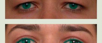
In what cases is umbilicoplasty performed?
There are no medical indications for navel surgery, but the operation is performed at the client’s request in order to eliminate the patient’s psychological discomfort that arises due to the presence of an aesthetic defect on his body.
Indications for umbilicoplasty:
- elevated position of the navel (in relation to the anterior surface of the abdominal wall, the scar protrudes by 0.5-1.5 cm);
- cicatricial deformities of the umbilical region - occur against the background of mechanical damage to the skin or an unsuccessful piercing procedure, followed by rupture of the skin and wearing heavy jewelry, which causes sagging of soft tissues and the formation of scars;
- T-shaped scar, which occurs as a result of overhanging (ptosis) of the skin over the vertical depression of the navel;
- deep funnel (more than 0.5 cm) – complicates hygienic care and promotes the accumulation of dirt;
- a spherical formation covered with skin, which is located inside the umbilical funnel (gives it an unaesthetic appearance);
- wide base of the navel - stretching of the tissues of the umbilical ring, which occurs under the influence of gradually increased intra-abdominal pressure during pregnancy and childbirth;
- incorrect location of the navel (violates the visual symmetry of the anterior abdominal wall);
- restoration of a lost navel, the loss of which was caused by surgical intervention in the abdominal cavity;
- umbilical hernia.
Umbilical hernia in adults: symptoms and after what it occurs
With a pathology such as an umbilical hernia in adults, the symptoms may vary - in some the pain syndrome is significantly pronounced, while in others it is completely absent; the degree of protrusion and the severity of the general condition also differ. The severity of the clinical picture will directly depend on how large the umbilical hernia is in adults. Symptoms in most cases include the following:
- A protrusion in the navel area is the main symptom of an umbilical hernia; this is especially noticeable in the photo when there are “before” and “after” photos. At first, it may be barely noticeable, painless and not alarm the patient in any way. At this stage, the patient can reduce the hernia on his own; often it can be reduced by itself in a lying position. As the disease progresses, the size of the hernia increases.
- A significant increase in protrusion and the inability to reduce it - such a symptom may indicate the formation of adhesions.
- An increase in the size of the hernia during a long stay in an upright position, coughing, sneezing, straining, after physical exertion, overeating.
- Pain - this symptom is not observed in all patients with a hernia. May occur after physical activity. Often indicates the formation of adhesions, or strangulation of a hernia.
- Sometimes the skin over the hernia becomes so thin that you can see the intestinal loops (photo on the right) and feel peristalsis.
- Dyspeptic disorders (constipation, belching, nausea) - such symptoms are observed very rarely, often already with the development of adhesions.
To prevent the development of an umbilical hernia, you need to know when it occurs in order to avoid surgery to remove it. So, a hernia can occur under the influence of the following factors:
- An umbilical hernia often occurs in adults after pregnancy (photo of a hernia in pregnant women on the right) - this is facilitated by multiple births, polyhydramnios, late pregnancy, and complications during pregnancy.
- Obesity, or rapid weight loss.
- Diseases accompanied by increased intra-abdominal pressure.
- Tendency to constipation.
- Impaired collagen synthesis, which leads to connective tissue weakness.
- An umbilical hernia often occurs in adults after abdominal trauma or surgery.
- Sedentary lifestyle.
Treatment of umbilical hernia in adults. Treatment by surgery
An umbilical hernia in adults, the treatment of which is delayed, can lead to serious complications, including strangulated hernia, peritonitis, intestinal obstruction, and necrosis of the strangulated organ.
Patients often wonder whether an umbilical hernia in adults can go away on its own. Treatment, according to experts, is not necessary only in children under 5 years of age, since they have a high probability of self-reduction of the hernia. In adults, treatment of an umbilical hernia is mandatory! The only effective treatment for umbilical hernia in adults is surgical removal (surgery). You can often hear or read on the Internet that treatment of an umbilical hernia in adults is possible not only by removal, but also by reduction, massage, and bandage. However, such methods are not effective and can have serious consequences.
How is an umbilical hernia removed in adults? Treatment (surgery) can be carried out in several ways - tension hernioplasty, non-tension hernioplasty, laparoscopic surgery. If it is a strangulated umbilical hernia in adults, treatment (surgery) is performed urgently, but if there is no strangulation, the operation is performed as planned. Which method will be used to perform the operation depends on each individual case - this is decided by the surgeon after a complete examination of the patient.
Buttock enlargement is no longer in fashion! Now everyone is doing navel surgery (6 photos)
Author: Eva Tushenkina
23 February 2020 15:10
Community: Facts
Tags: appearance plastic surgery plastic surgery plastic surgery plastic surgeon navel improvement
12369
6
According to data for 2020, Americans and American women spent $16 billion on improving their appearance through plastic surgery. Surgeons say that the fastest growing trend today is umbilical plastic surgery, or umbilicoplasty. Demand for it has doubled over the past few years! Patients ask for neat belly buttons, like those of famous actresses and models. A special excitement begins in the spring, when the fair sex thinks about a summer vacation.
0
Source:
See all photos in the gallery
According to plastic surgeon Matthew Shulman, the demand for navel surgery has increased thanks to social networks and the availability of any information. Another doctor, Dr. Thomas Sterry, believes that the navel is an important part of our anatomy from a cosmetic medicine point of view. And if you go to the beach and your belly button is not perfect, then you are unlikely to look sexy.
0
Source:
Dr. Shulman says the ideal belly button is a vertically oriented oval, which visually lengthens the belly and makes it appear flatter. “When it comes to belly button jobs, people bring pictures from Victoria Secret catalogs,” he says.
0
Source:
Also popular among Shulman’s patients are photographs of actress Jessica Simpson from 2005 (when she played in the film “The Dukes of Hazzard”) and model Emily Ratajkowski.
0
Source:
And sometimes they even ask for a “hidden” or “sewn up” navel. Yes, there is such a fashion now.
0
Source:
Very often, victims of umbilical piercing turn to surgeons. “I call it the Britney Spears effect,” says Dr. Shulman. “Those young girls who got their belly buttons pierced in the 90s are now grown, they are about 35 years old, and often their belly buttons look very sloppy due to weight gain or after childbirth.”
0
Source:
The operation is laparoscopic and is performed through small incisions made around the navel area. It is done under local anesthesia. Takes 30-40 minutes. According to Dr. Sterry, he performs at least 20 umbilicoplasties a year - and five years ago this figure was half that.
Source: - translated specifically for
Related links:
- Japanese cosplayer spent $64,000 on a total makeover of her appearance
- The Iranian woman was eager to turn into Angelina Jolie
- Mother did not recognize her son after plastic surgery performed on him
- The Twins Paid $1 Million to Look Like Brad Pitt, But They Didn't Work Out
- 63-year-old Kim Basinger scared fans with her frozen face after plastic surgery
subscribe to the “Facts” community
Tags: appearance plastic surgery plastic surgery plastic surgery plastic surgeon navel improvement
Did you like the post? Support Chips, click:
108 60 48
Liked
48 0
55
Partner news
Removal of an umbilical hernia in adults, photo of an umbilical hernia
Removal of an umbilical hernia in adults is performed under local or epidural anesthesia.
Hernioplasty (hernia repair) can be performed either traditionally surgically or laparoscopically. Traditional removal of an umbilical hernia in adults involves making an incision in the skin and subcutaneous tissue. Then the hernia is either reduced inside the abdominal cavity (if the size of the defect is small), or it is completely removed. The next step is to strengthen the umbilical ring. For this, either the patient’s own tissues (tension hernioplasty) or synthetic materials (non-tension hernioplasty) are used. Removal of an umbilical hernia in adults using a non-tension method is more effective - it allows you to eliminate even large hernias and is characterized by a low likelihood of relapse. Recently, laparoscopic removal has become increasingly popular (photo above). It does not require an incision, is characterized by a short rehabilitation period, and a low risk of complications and relapses.
They will help to recognize such a pathology as an umbilical hernia in adults - photo. They can be found on various medical websites, as well as in patient reviews. The protrusion of the navel immediately catches the eye - in some patients it is more pronounced, in others less. The symptoms mentioned by the patient, palpation and ultrasound help the doctor confirm the diagnosis of a hernia. What does a strangulated umbilical hernia look like in adults? Photos will not help determine whether the hernia is strangulated or not. The differences between a strangulated hernia are symptoms such as acute pain, dyspeptic disorders, or disorders of intestinal function. Photos of umbilical hernia in men and women are above.