Scars appear on the human body after injuries (scratches, cuts, burns, etc.), skin diseases (acne, boils, smallpox, etc.) and surgical operations. A scar is a consequence of the replacement of a skin defect with connective tissue. There are different types of scars that differ in structure and appearance.
Some defects are easier to get rid of, others more difficult. To speed up scar healing and prevent complications, the patient should know how to care for it after surgery or injury.
What do we understand by the concept of scar?
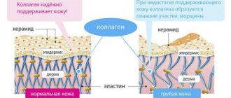
A scar is a mark on the skin after it has healed. Skin healing involves replacing normal skin with connective tissue, which is the final stage of skin restoration. Connective tissue is significantly different from normal skin. She has much lower regenerative abilities. For example, hair follicles and sweat glands are not restored at the site of the scar.
And in appearance, scar tissue differs significantly from healthy skin: scars stand out against the background of normal skin in color and texture. When the skin is damaged, a “tear repair” mechanism is triggered, which helps the wound heal. If the wound is superficial, then no mark remains on the skin. When the deep layers of the skin are damaged, scars appear at the site of the wound.
Recommendations
Experienced farmers and veterinarians know what needs to be done to protect the livestock and not incur financial losses.
- Avoid grazing cows in fields sown with legumes and cereals.
- Don't switch abruptly from one type of food to another. It is better to stretch this process over a couple of weeks.
- Give young animals hay before putting them out to pasture to reduce their appetite and thereby prevent them from overeating on grass.
- If possible, install magnetic planes in barns. They will protect you from accidentally getting metal objects into your food.
- Monitor the absence of foreign debris in grazing areas. The grass may contain nails, glass, pieces of metal cans and other dangerous objects. Since the cow's mouth is equipped with papillae directed inside the body, if garbage gets in with food, the animal is no longer able to spit it out.
Scar classification
Scars are of the following types:
- Physiological or normotrophic scar. This scar is located at the surface of the skin. That is, it does not rise above it. After healing, the wound becomes almost invisible and after a few years it is impossible to find it. In this way, minor wounds and scratches, burns and abrasions, or skin incisions made by a competent surgeon during surgical interventions heal. Experienced surgeons try to make incisions in natural skin folds. Incisions made in such places make scars almost invisible.
- Atrophic scar. Formed as a result of extensive damage to the skin and subcutaneous fat. This scar is located below the skin level. That is, it seems a little pulled inward. Atrophic scars often form after acne, chickenpox, furunculosis, or as a result of electrical trauma.
- Hypertrophic or pathological scar. Scars of this type are considered a serious aesthetic defect. They are rough, dense, and rise above the surface of the skin. Sometimes peeling, redness or trophic ulcers are noticed on such scars.
- Keloid scar. This type of scar is the most unfavorable outcome of wound healing. Outwardly, it looks like a tumor, rising greatly above the skin. It has a bright pink or bluish color, uneven texture and a certain density. Its base is always larger than the area of the wound itself. Causes pain, itching and burning. To date, its etiology is not completely clear. The main factor in its development is considered to be a hereditary predisposition to the formation of keloid tissue, as well as a specific location of skin damage (sternum, pubic area, ears).
Let us dwell in more detail on hypertrophied and keloid scars.
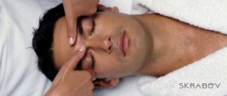
Surgery
At the moment, there are many methods of surgical excision of scars. When choosing a specific method, the specialist pays attention to the density, size, and type of scars. What is important is the condition of the skin surrounding the scar, and the position of the scar relative to other landmarks of the human body.
Much attention is paid to complex scars that arise as a result of surgical operations, after serious burns, and deep injuries. What features does the operation have:
- Small scars that protrude slightly above the skin level are excised with a scalpel or blade parallel to the skin. Here it is very important not to cut off too much, to avoid this, the raised part of the scar is marked. Uneven boundaries between the removed scar and healthy skin are corrected using electrosurgical instruments. The healing of such a wound occurs by secondary intention, that is, it is not sutured.
- Another common method of scar excision is spindle-shaped or elliptical excision. Typically used to treat scars that run across the natural lines of a person's skin. An incision is made across the scar, so that the new one coincides with the natural line, the edges are excised slightly and finished with sutures. A temporary suture is applied if the original scar was long enough, and then the operation requires several such approaches to completely remove the old scar. The result is a new scar, made across the old one, so that it matches the natural lines on the skin and does not stand out. Its healing occurs under correction and supervision, so that the result is a neat, almost invisible defect. If the scar was initially hypertrophic, then it is first excised to level it with the level of the skin, so the entire course of correction can sometimes take six months or a year. Patients must be prepared for this.
- Skin grafting and flap plastic surgery are popular. Thanks to these methods, it is possible to move a serious scar from a cosmetic area to less noticeable places on the human body. I often use it for burns and keloid scars. The scar is excised and the area is covered with skin from the donor site. As a result, instead of a scar, almost invisible small scars remain. This result is considered acceptable.
- In dentistry, excision and plastic surgery of scars on the oral mucosa are also used. Sometimes, atypical scars are found on the mucous membrane, and they are excised because they interfere with prosthetics. Among these operations on the oral mucosa, various types of plastic surgery and transplantation are also used.
In addition to these techniques, there are other types of plastic surgery, for example, W-plasty or Z-plasty and others, it all depends on the type of scar. But all methods are effective, and these are the results you can achieve if you approach the problem responsibly:
In any case, surgical excision or scar plastic surgery are radical techniques. Doctors advise resorting to them if the problem is really serious and other types of therapy do not help.
Hypertrophic scars
The term "hypertrophic" consists of two parts: "hyper", meaning "excessive", and the Greek word trophos ("nutrition").
Hypertrophic scars are formed as a result of excessive formation of connective tissue during the wound healing process. The intercellular matrix is responsible for the formation of all scars. After skin damage, myofibroblasts (smooth muscle cells that can transform into fibroblasts) send collagen, elastin and other proteins to the wound site, which leads to the formation of scars.
If the normal wound healing process is accompanied by inflammatory processes or other unfavorable factors, then a very intensive process of collagen formation begins. As a result, this leads to excessive accumulation of proteins, the development of skin fibrosis, which leads to the formation of hypertrophic and keloid scars.
The appearance of a scar is good for the body. After all, the wound is closed, which means that the danger of infection or blood loss has passed. However, the aesthetic unattractiveness of such scars causes psychological discomfort.
Cow rumen does not work - folk remedies
02/06/2020 Category: Recipes from the peopleAuthor: Dmitry
Appetite in cattle is an indicator of health. Its absence alarms the owner, because it can be a harbinger of many diseases of various origins. Often only a specialist can help determine the cause. But sometimes his help is urgently impossible. The livestock breeder needs to master basic actions that will provide first aid to the cow. This publication will discuss stomach diseases.
In cattle, problems with the functioning of the stomach are noticed quite often. In some cases this is due to the human factor. The owner gives the cow low-quality food, heavy, too cold or hot food. The cow, in turn, is sometimes not too picky and swallows food that is harmful to itself, and often foreign objects. Harmful to the cow's stomach:
- consumption of poorly chopped root vegetables, corn, pumpkin and other types;
- foreign objects, including nails and stones;
- starvation, when the animal begins to randomly and greedily swallow food provided belatedly;
- fright and, as a result, stomach spasm (a phenomenon often observed in calves), the esophagus narrows greatly and becomes.
Based on these prerequisites, the appropriate recommendations for feeding pets should be followed. In particular, give the cow only fresh food, well crushed to avoid stomach cramps.
Vegetables can be grated or chopped. Corn cobs need to be cleaned. Avoid long breaks between meals.
In some cases, the course of the disease is facilitated by paralysis of the esophagus. This occurs due to injury to the mucous membrane of the esophagus and the muscle wall. In addition, a tumor may grow there due to tuberculosis or leukemia. Inflammation of the brain during infections such as rabies also leads to paralysis.
The disease will not take long to appear; signs appear immediately if the stomach does not work:
- belching disappears and the chewing reflex disappears;
- refusal to eat and open mouth;
- the cow begins to chew the void so that foam comes out;
- nervous tail wagging;
- the cough reflex is triggered;
- breathing is heavy, with shortness of breath.
If food or an object gets stuck in the area of the larynx, it will be visually noticeable. Then the cow gags and tries to push him out. She often succeeds.
When the blockage drops lower, the animal's anxiety increases. When there is a partial blockage, gases regurgitate through the esophagus, and water can reach the stomach. But the digestion process is still disrupted, and the cow’s stomach becomes irreversibly damaged.
An erect stomach not only causes discomfort to the cow, but can also lead to its death. Therefore, treatment should follow immediately after symptoms are detected. Since help must come quickly, many livestock farmers have questions about what can be done at home if the cow has a stomach problem.
First, you should carefully examine the animal. Perhaps the illness occurred due to a foreign object that can be reached. Do this:
- The cow should be properly tied and should not push or move.
- Wrap your hand in a rag up to your shoulder so as not to damage it.
- To prevent the animal from closing its mouth, insert a wedge between the teeth.
- An assistant should place the object between the teeth if the wedge suddenly falls out.
- pour in a little vegetable oil to make the probe glide smoothly.
- you can pour in a little water, up to three liters, to push the object through.
Sometimes, at home, you can force a cow to stretch out its tongue, irritate it in every possible way in order to vomit. The hope is that she will be able to spit out the foreign body.
In addition, antispasmodics can be injected intramuscularly under the skin. If you don’t have a probe at hand, a rubber hose with round edges will do. It is advisable that the diameter does not exceed three and a half centimeters. Hard objects such as sticks should not be used, as they can injure the esophagus.
Sometimes the victim is a calf. Newborn babies often experience bloating. It is not yet developed enough, only the food gutter works and the stomach stops systematically.
If you don’t have the necessary medications in your first aid kit or you can’t physically push an object or a lump of food through, there are still options for what to do at home if the cow has a stomach problem. These are folk recipes:
source
Gastric congestion in a cow is a problem that requires a quick solution. Untimely assistance can have a detrimental effect on the subsequent life of the animal. Let's figure out what to do if a cow has a stomach ache and how to treat this condition.
Stopping the work of the stomach is a consequence of one of the following reasons:
- Eating food that has been chopped into large fractions. The gastrointestinal tract cannot process insufficiently detailed vegetables (corn, carrots, turnips, pumpkin and others), which leads to stagnation.
- Insufficient crushing of food by the animals themselves. A hungry animal, when actively and randomly eating food, can swallow it almost entirely, which is why the rumen subsequently does not work.
- Entry of a foreign body. If large objects, such as bones or stones, enter the cow's esophagus along with food, injury and complete blockage occurs, as a result of which the digestive tract cannot continue to function properly.
- Stress. This is one of the most common reasons why a calf's stomach does not work, especially after calving. An animal that has experienced fright or severe emotional shock may experience a spasm of the digestive tract.
- Tumor in the esophagus. One of the probable symptoms of tuberculosis and leukemia requires immediate identification.
- Paralysis of the esophagus. It can occur after injury to mucous membranes or muscles, or as a result of rabies infection.
The following signs indicate gastric congestion in a cow:
- the cow does not eat, does not show appetite;
- the chewing reflex disappears;
- the animal stops burping;
- frequent movements of the jaws appear;
- productivity decreases;
- general malaise is expressed;
- the animal begins to breathe heavily with accompanying shortness of breath;
The animal is provided with rest and a small diet with a reduction in the amount of food and the removal of low-quality food, after which therapy begins. There are several methods for treating gastric congestion. These include: manipulations to remove a foreign object, conservative drug treatment, surgery and traditional methods.
If the cause of gastric stoppage is the entry of a foreign object, it must be removed.
Drug treatment in the form of antispasmodics can be used if there is a need to remove a foreign body. In other cases, the veterinarian may prescribe Carbacholine injections under the skin or taking medications for the cardiovascular system with glucose.
If previous treatment methods do not produce results, surgical intervention is resorted to. The cow undergoes an esophagotomy, an operation to cut open the esophagus to examine or remove foreign objects.
Gastric arrest in a cow can be treated with traditional methods:
- Add 100 g of yeast to 200 g of warm water, leave for 30 minutes, then add 200 g of vodka and 100 g of granulated sugar and mix thoroughly until smooth. The animal should take the infusion for several days, 2 times a day.
- A decoction is made and infused from St. John's wort, flax seeds and yarrow. Apply 2 times a day until the digestive tract is restored.
- Mix 0.5 liters of water with hellebore tincture and allow to be taken orally.
- The stomach is washed with a solution of baking soda or table salt.
To prevent gastric arrest, it is important to take preventive measures:
- The cow should be given high-quality feed.
- Vegetables must be thoroughly chopped.
- You should not leave an animal hungry for a long period of time.
- Maintenance and milking must be carried out in accordance with accepted standards.
- The animal should be checked for diseases in a timely manner and routine tests should be carried out.
Gastric arrest in a cow can be successfully treated if you promptly pay attention to the symptoms and unusual behavior of the animal.
Proper therapy will start the work of the gastrointestinal tract, and prevention will help prevent the occurrence of stagnation in the future. source
Gastric arrest in a calf is a dangerous condition that can be fatal if the animal is not treated in a timely manner. The farmer needs to know how this problem manifests itself, as well as what to do and how to properly start the stomach.
Gastric arrest in a calf
Most often, the stomach in calves stops if there is a blockage in the esophagus. This can happen when a hungry animal greedily ingests large pieces of food, such as root vegetables or corn on the cob. A large piece of vegetable ends up in the esophagus, where it can get stuck, never moving into the rumen.
This situation is dangerous for the calf - after the stomach stops, gases begin to accumulate in it. As a result, it increases in volume and puts pressure on the diaphragm. This makes it difficult for the cow to breathe. If help is not provided in time, death from asphyxia can occur in a little more than a day. That is why it is important to detect the problem in time and help the animal.
Let's look at the most common reasons for rumen failure in calves:
- Ingestion of foreign objects (rags, wood chips, nails, stones).
- Ingestion of large fragments of food (beets, potato tubers).
- The animal is frightened while eating food - in such a situation, a spasm of the esophagus may occur, its lumen narrows, so the food does not pass further.
Attention! Narrowing of the esophageal tube occurs not only due to fear - sometimes it happens with serious illnesses in the cow, for example, leukemia, tuberculosis or problems with the nervous system, as well as with tumor processes in the neck or chest area.
Stagnation in the rumen of calves often occurs when they are switched to solid feed. A provocateur of this condition can be starvation - if a cow constantly receives insufficient food, and then suddenly overeats, the stomach, out of habit, cannot cope with the heavy load and stops.
The stagnation process in the rumen is manifested by changes in the behavior of the animal. If it is caused by a blockage of the esophagus, you may notice the following:
- The calf stretches its neck and tries to burp a lump.
- The mouth is slightly open.
- There is no chewing gum.
- The cow makes idle chewing movements.
- The saliva produced is predominantly foamy.
- The animal is worried, scared, shaking its head.
- When the lumen of the esophagus is completely blocked, the animal does not eat or drink.
- When the esophagus is partially blocked, the calf tries to eat, but due to difficulty swallowing, it soon stops, but accepts drinks and liquid food.
Stomach failure leads to the accumulation of gases in the rumen, as a result of which anxiety only increases. Subsequently, the clinical picture changes:
- Shortness of breath appears.
- Breathing becomes shallow.
- Abdominal bloating is observed.
- A cough appears.
Attention! Symptoms of stagnation in the rumen cannot be ignored. If you do not help the calf at this stage, it will die.
If you suspect a stagnant process in the rumen, you must urgently invite a veterinarian to find out the cause of this condition. If it is discovered that a foreign body is stuck in the esophagus, it must be removed immediately. This can be done in several ways - try to remove it manually through the mouth or push it into the scar using a probe. The choice of method depends on where exactly the foreign body is located - in the cervical esophagus or lower, in the thoracic.
Removing a closely located object from the esophagus is carried out as follows. First, the animal is given about 100-150 ml of vegetable oil to drink. If possible, the esophagus is irrigated with a solution of novocaine for pain relief. To relieve spasms, Spazmalgon or other antispasmodics are used intramuscularly. The calf is fixed, a wedge is inserted into the oral cavity between the teeth so that the mouth does not close. The veterinarian wraps the hand in a towel to prevent injury. The limb is inserted into the esophagus and the stuck object is carefully removed from it.
If the foreign body is closer to the scar, so that it is not possible to reach it with your hand, then they resort to using an elastic probe. It is slowly inserted into the esophagus and the bolus of food is pushed into the rumen.
Attention! It is forbidden to use sticks or rods instead of a probe - there is a high risk of perforation of the esophagus.
If the calf has a bloated belly, it is necessary to release the gases from the rumen. The veterinarian does this. He pierces the peritoneum in the side of the body and inserts a cartridge case into it. Air will escape through this hole. Immediately after the procedure the animal will feel better.
Starting the stomach includes following a diet on the first day after all the above-mentioned manipulations. The calf is given plenty of water to drink, but not fed. On the second day, food is offered in the form of liquid mash. As the functioning of the digestive tract improves, grass, hay, and root vegetables in crushed form are gradually introduced.
Folk remedies help to get your stomach started. Most of them are not suitable for calves, such as mixtures with vodka, but there are others that are applicable to young animals. Calves are allowed to drink:
- Mineral water.
- Brine (cucumber or sauerkraut).
- Lactic acid.
Farmers note that herbal decoctions give good results when starting the stomach.
Take any raw material - St. John's wort or chamomile flowers in an amount of 25 grams, brew with a liter of boiling water. Simmer on the stove over low heat for 15 minutes. The product must infuse. Then it is filtered. The calf should drink 350-400 ml of herbal decoction in the morning and evening.
Hellebore tincture, diluted with water, is also used to treat congestion in the stomach. It is sold at the pharmacy. The entire volume of the bottle is combined with half a liter of boiled water. The calf is given 100 ml of this medicine in the morning and evening.
Reference. If no action helps solve the problem - to start the calf's stomach, then surgery will be required. It is performed in a hospital under general anesthesia. This method is also used if a tumor of the esophagus is detected.
It is better to try to prevent any problem. The same rule applies to gastric arrest in a calf. To prevent this from happening, you need to carefully check the food supply for the presence of foreign bodies and debris. The farmer must keep the stall clean. Food for calves must be thoroughly chopped - cut into small pieces or grated. Animals should not be allowed to starve, after which they usually eat hastily.
Gastric arrest is dangerous for calves and adults and can lead to the death of the animal. The farmer needs to be careful and observe the appetite and behavior of the cows. If stagnation is detected in the rumen, you should immediately invite a veterinary service worker.
source
Many novice farmers are interested in the question of what to do if a cow’s stomach is stuck. Note that there are quite a few reasons for the occurrence of such a condition in cattle. This pathology can be provoked by various unfavorable endo- and exofactors. Therefore, we will consider the main causes of this unpleasant disease, we will present effective treatment methods that will help to improve the cow’s stomach
Unlike other animals, esophageal blockage is very common in cows. Cattle suffer from tympany, which occurs due to excessive accumulation of gases in the rumen.
As a rule, disturbances in the functioning of the digestive tract organs arise due to unbalanced feeding and low-quality, low-grade feed. If a cow has a stomach problem, one of the reasons is the presence in the animal’s diet of poorly chopped corn cobs or, for example, large pieces of pumpkin.
Considering that cows are characterized by mineral starvation, which is caused by an insufficient supply of useful macro- and microelements, vitamins, and amino acids with feed, animals can eat foreign objects. The stomach of cattle often stops due to the presence of foreign bodies in the gastrointestinal tract.
When swallowing objects whose diameter is larger than the lumen of the esophagus, the organ reflexively contracts, and saliva is swallowed. This is a kind of protective reaction that promotes the pushing and passage of an object along the esophagus. At the same time, pressure on the walls of the organ increases, acute inflammation and swelling develop. Later, necrosis appears and perforation of the esophageal wall occurs. If the tissues become infected, an infection develops that spreads to the pleural cavity and mediastinum. Aspiration pneumonia may develop.
Important! Exhausted, weakened, and hungry animals are at risk. The cow, experiencing severe hunger, begins to quickly absorb food. A cow's stomach can stop if the walls of the digestive tract are injured by sharp objects.
Gastric arrest in cattle can be caused by:
- a sudden change in diet;
- unbalanced feeding;
- severe stress, fear;
- development of neoplasms in the gastrointestinal tract;
- ingestion of bones, tendons, large solid objects;
- congenital anomalies of the digestive tract.
Rotten, fermented feed, rotting hay can also provoke tympany, flatulence, and gastric arrest in farm animals.
Blockage of the esophagus in cows can occur from severe fright or a sudden spasm of the organ. There is also an unnatural narrowing or paralysis of the esophagus. This pathology is caused by injury to the mucous membrane of the esophagus and muscle walls. It may occur if the mediastinum is compressed by a developing tumor, occasionally with tuberculosis, leukemia due to enlarged lymph nodes.
Paralysis of the esophagus in cattle can develop in the event of compression of the organ itself (peripheral paralysis), inflammation of the brain (central paralysis), or neurotropic infections such as rabies. Infectious, parasitic diseases of cows can also cause this pathology in cattle.
This pathology is also observed in small calves at the age of 1 month. Newborn babies suffer from bloating and gastric constipation. This is due to the fact that the calf’s gastrointestinal tract is still poorly developed, and only the food gutter works. This usually goes away by 2 months, after the calf is switched to dry food. Together with them, beneficial bacteria enter the esophagus, which normalize the functioning of the gastrointestinal tract.
Your veterinarian will advise you on how to cure this pathology in small calves. Without experience and the necessary knowledge, self-medication can only aggravate the situation.
As a rule, this pathology develops gradually. In the initial stages of the disease, signs appear subtle.
Important! Blockage of the esophagus in cows can be complete or incomplete.
When the blockage is complete, cows become timid, show anxiety, constantly moo, stretch their necks forward, lower their heads, and nervously beat their tails. In sick animals, the ruminant reflex disappears. Appetite decreases or is completely absent. The oral cavity is constantly open. When trying to swallow saliva, a foamy, viscous liquid flows out of the mouth. Breathing is rapid, shortness of breath appears. The cow constantly tries to burp and coughs. If a cow's stomach stops, defecation is disrupted, and there may be prolonged constipation.
In case of incomplete blockage, in the area of the neck, jugular groove, you can notice a dense spherical compaction, the presence of a foreign object upon palpation. Animals burp air, can take food in small portions, and drink water. Outwardly, cattle do not show signs of severe anxiety, but over time the stomach becomes upset and the cow’s condition worsens.
Severe concern is noted when large foreign objects enter the thoracic esophagus.
In case of gastric arrest in cattle, in order to prevent suffocation, it is necessary to provide assistance to the sick animal as soon as possible. If this pathology is not treated, the cow can die within a day. If a cow has a stomach problem, a veterinarian will select and advise treatment methods for this pathology. Treatment depends on the form of the disease and the location of foreign objects.
Considering that the main cause of gastric arrest is the presence of a foreign body in the esophagus, it must be removed as soon as possible. All actions should be performed in a certain sequence together with an assistant:
- Secure the cow well, install a wedge between the molars to secure the jaw. If it accidentally falls out, assistants should quickly insert something hard between the teeth. It is best to use a probe.
- To avoid injury, wrap your arm from shoulder to hand with a towel.
You can remove a foreign object in another way. Wrap your hands around the cow's neck and place them in the area of the jugular groove. Perform movements towards the animal's head. Before the procedure, veterinarians recommend pouring 90–110 ml of any vegetable oil into the cow’s throat. You can provoke a gag reflex in a cow by gently pulling out her tongue. In this case, the foreign object should come out spontaneously.
If a cow's stomach is stuck due to a blockage in the cervical and thoracic parts of the esophagus, we recommend using an elastic probe, preferably a large caliber (35 mm), to pass a foreign object. It should move along the esophagus. To facilitate the procedure, you need to introduce a glass of sunflower oil. This will make it easier to move the item forward.
The pushing procedure must be carried out very carefully, gradually increasing the pressure of the probe. Otherwise, strong shocks can cause perforation of the esophagus.
Advice! Experienced farmers and livestock breeders recommend pouring 2-3 liters of warm water into the esophagus or pumping air at the same time as inserting the probe. Under pressure, the walls of the organ expand, and it is much easier to push through a foreign body.
If a cow's rumen is swollen, the symptoms of tympany must be relieved before removing the blockage of the esophagus. The scar is pierced with a trocar. Without experience, in order not to harm the cow, entrust this procedure to a veterinarian.
If you did everything correctly, but the methods described above did not lead to the desired result, a surgical operation will be performed - esophagotomy.
To relieve pain and spasms, animals are given analgesics. For example, Spazmolgon, which is administered intramuscularly in a dose of 1 ml. You can use No-shpu, Drotaverine, Oxytetracycline, Sulfocamphocaine, cardiac glycosides.
If a cow's stomach becomes upset due to poor-quality feed, this means that the fore-ventricles have stopped working. Knead the area of the hungry pit on the left with your fist to stimulate the stomach.
Starting the stomach in cows can also be done using alternative medicine. Dissolve 100 g of yeast in 250 ml of warm water. Leave to soak for 25-30 minutes. Pour 200 ml of vodka into a separate container, add 100 g of sugar and yeast diluted in water. In total, you should get 1 liter of tincture. If necessary, add water to the required volume. The prepared solution is poured into the cow's throat twice a day for several days.
If a cow’s stomach does not work, experienced livestock breeders advise using hellebore tincture, which is diluted in a half-liter bottle of water and poured into the animal’s mouth.
When the stomach stops, the cow can be given a decoction of yarrow, chamomile, St. John's wort, and flax seed as a drink. 20–25 g of medicinal raw materials per liter of water is enough. The tincture is poured down the throat of the animal twice a day.
It is recommended to give a month-old calf pickle from cucumbers, cabbage, and tomatoes. If the calf's stomach does not work, give it mineral water or add lactic acid to its drink.
source
Clogging of the esophagus in livestock is an acute pathology, which is manifested by partial or complete blocking of the lumen of the stomach with large pieces of food or foreign objects. This condition requires urgent treatment. If cattle are left untreated, it will be fatal.
The specificity of blockage of the esophagus in a cow is that the signs of this pathology appear abruptly. The animal experiences severe discomfort. The problem can be identified by the following symptoms:
- the cow looks scared;
- loss of appetite;
- jaw open;
- the animal often performs swallowing movements;
- there is no chewing gum;
- chewing movements occur idle;
- no belching;
- the cow shakes her head;
- severe salivation, sometimes in the form of foam.
When the cow's esophagus is completely clogged, the stomach stops. Gases accumulate in the rumen, constipation appears, and the individual’s well-being noticeably worsens.
Over time, other signs appear:
- The individual is nervous and cannot stand still.
- There is heaviness in breathing.
- The cow tries to hit itself in the stomach with its hoof.
- Swings its tail.
- Cough and shortness of breath appear.
- The mucous membranes acquire a pale tint.
If the blockage is partial, the cow will try to continue to eat, but the attempts will not show the desired result, since swallowing is complicated. However, the animal can drink and eat liquid food.
If the pathology is detected in time, the blockage is removed and the animal recovers. Complete blockage of the esophagus and failure to provide timely assistance (within 1-3 hours) will lead to respiratory arrest and death of the livestock. With prolonged partial blockage, inflammation may occur, as well as necrosis and rupture of the gastrointestinal tract membrane. In severe conditions, aspiration pneumonia develops.
There are many reasons why the stomach becomes clogged and stops working. The problem appears due to non-compliance with the rules of keeping livestock (diet, size of food, cleaning in the pen.). Pathology develops for the following reasons:
- Ingestion of a foreign body (stone, glass, metal). Objects enter the esophagus simultaneously with food.
- Failure to comply with the diet leads to overeating.
- Supplying food in the form of vegetables in fairly large pieces.
- A long gap between feedings will cause the cow to quickly eat food, swallowing it and not chewing it. The food will get stuck and will not be able to pass into the stomach.
- Blockage forms in the presence of cuts and spasms, as there is a failure of gastrointestinal motility. The food stagnates and the animal is unable to swallow it.
Also, individuals experience mineral hunger (lack of vitamins and minerals), resulting in the habit of swallowing foreign objects.
- Complete - the gastrointestinal tract gap is completely closed.
- Partial - the lumen is half closed, for example, when swallowing a flat object.
Complete blockage occurs due to rapid eating of regular food or large feed. Partial appears when involuntary swallowing of foreign objects that are oval or elongated in shape. They become horizontal in the stomach and do not pass further. If measures are not taken in time, an inflammatory process in the esophagus may develop, as well as swelling at the site of the foreign body or tumor.
Methods such as inspection, palpation, probing, esophagoscopy and fluoroscopy will help to make a correct diagnosis in cattle. Each type has its own characteristics and is carried out exclusively by a veterinarian. The studies are characterized as follows:
- Inspection. This method helps to see the difficulties in the passage of food. The veterinarian examines part of the left jugular recess, paying attention to wave-like movements along it. The area is fine if water and food pass normally through the gastrointestinal tract. If its size is increased, this indicates the presence of inflammatory edema, a plug of foreign objects, a rupture and other neoplasms.
- Palpation. The diagnosis is performed with the left hand, holding the abdominal part of the esophagus. With the right hand they probe the cervical part along the jugular groove. This method helps to determine the presence of diseases of the gastrointestinal tract and other tissues, as well as to identify the presence of foreign objects.
- Probing. You can reduce gases in the intestines and remove feed mass using this method. This method helps to quickly determine the cause of the disease, after which the veterinarian decides how and with what to treat the animal. This method is also actively used for artificial nutrition, gastrography, and gastrotonometry. The probe is selected depending on the age and weight of the cow. Before the procedure, the equipment is disinfected.
- Esophagoscopy. This is one of the effective ways to study the esophagus. The procedure has two directions - therapeutic and diagnostic. The first is addressed in severe cases. For example, if a cow has swallowed a foreign object, as well as with burns and inflammation. This method is used to introduce the necessary drugs into the intestines or apply them to the walls. By diagnosing the esophagus by esophagoscopy (the second method), it is possible to qualitatively examine the animal. Thanks to this method, the veterinarian will be able to take a particle of the gastrointestinal mucosa for analysis. Thus, the disease can be detected at an early stage and treatment can be started in a timely manner.
- X-ray. Veterinarians use X-rays to examine and detect various diseases in cattle. This method is considered auxiliary for establishing a diagnosis, using other methods. Transillumination should always be performed simultaneously with other studies. This is the only way to accurately diagnose and prescribe treatment.
The esophagus is clogged and the cow’s stomach is stuck, what should I do? A common question that attracts the attention of farmers.
If a cow has swallowed a stone, large vegetable or any other object, it is removed by hand, having previously tied the animal tightly to a tree. To prevent the mouth from closing, a wedge is placed between the teeth. A person's hand is tied with cloth. However, it would be safer to use an umbrella if such a problem occurs. It goes deep into the esophagus and pushes the object well. To ensure that the umbrella passes freely, the cattle are given vegetable oil. In addition, water is poured into the cow at the same time as the umbrella to improve the passage of the stuck body.
If the animal has rumen swelling, signs of tympania (bloating) are eliminated before treating the blockage. The scar should be pierced with a trocar. If the farmer does not have experience in this matter, it is better to contact a veterinarian so as not to harm the cow.
If the farmer has done everything correctly, but the cattle continues to feel unwell, surgical intervention in the form of esophagotomy (dissection of the walls of the gastrointestinal tract) is necessary. Pain can be relieved and the animal’s condition can be alleviated using analgesics. The cow is given Spazmalgon, which is administered intramuscularly in a dosage of 1 ml. No-shpa, Drotaverine, Oxytetracycline, Sulfocamphocaine and cardiac glycosides also reduce pain.
If your stomach is upset because of bad food, then your pancreas does not work. The farmer can massage the animal with his fist in the area of the hungry pit on the left. Thus, the stomach will be stimulated.
You can also cure an animal using folk recipes that are easy to prepare at home. To do this, just use one of the proposed options:
- Dissolve the package of yeast in a glass of warm water. The mixture is left for 30 minutes to swell. Next, take a glass of vodka and add 100 g of sugar to it. All components are mixed and mixed well. Add water to the prepared tincture to obtain 1 liter of product. The infusion is given to the cow 2 times a day (morning and evening) in a dose of 0.5 liters.
- To prepare hellebore tincture, use yarrow, St. John's wort and flax seeds. All components are taken in the amount of 25 g per glass of water. I infuse the herbs for 2-3 hours, and then the animal sings 3 times a day.
- If the cow is calving, it is prohibited to give her vodka. To do this, use herbal decoctions to stimulate the esophagus. Chamomile, yarrow and flax are suitable for treatment. A liter of hot water is poured into the greens (20-25 g). Then the broth is simmered over low heat for 30 minutes, infused for 1 hour and filtered. They water a cow with it 3 times a day.
- When the calf's stomach stops functioning, it is restarted with brine (cucumber or tomato).
The farmer needs to know that if there is a foreign body in the gastrointestinal tract and gas accumulation in the rumen, it is prohibited to give food and drink that can cause fermentation. First they fix the problem and only then start the stomach.
Every farmer must take responsibility for keeping cattle and monitor their health. It is necessary to feed cows strictly at the right time, thereby avoiding overfeeding and not allowing them to starve. The feed should be chopped and animals should not graze near potato, beet and cabbage piles.
To prevent foreign matter from entering the esophagus, the pasture and paddock are cleaned regularly and feeders are checked for large objects. In addition, vitamins, minerals and lick salt are added to the food. If the animal does not have enough nutrients, it will begin to lick walls, the ground, and also swallow foreign objects.
source
Reasons for the formation of hypertrophic scars
Hypertrophic scars occur as a result of surgery or severe burns. When wound healing is accompanied by inflammatory processes, decay products accumulate in the wound.
Hypertrophic scars are formed due to:
- deep burns, lacerations or bite wounds;
- lack of qualified medical care in the treatment of such wounds;
- infection or suppuration of the wound;
- permanent scar damage;
- location of the scar in active zones, which causes regular trauma;
- hereditary predisposition to active scarring.
What types of scars can plastic surgery remove?
In general, any scar or scar is the result of the process of replacing skin tissue at the site of damage with connective fibrous tissue. Only in some cases it can be a thin, barely noticeable pale strip, in others it can be a rough and large-scale scar, which causes not only moral discomfort, but also physical discomfort, because in some cases it may involve excessive tension of the skin. Such scars are also called keloids.
In general, there are several types of scars that can be removed by plastic surgery:
- small round scars or thin strips drawn in, which do not appear under the influence of external factors, but are the result of natural processes in the body. This includes stretch marks from pregnancy, acne scars, etc.;
- -with a smooth surface - can be white or pale pink, they appear either as a result of surgical interventions, or against the background of the independent development of fibrous tissue after minor injuries;
- keloids - they roughly rise above the surface of the skin, most often such scars have a dark burgundy color. In addition to external discomfort, they cause a person physical discomfort, because, firstly, due to the tension of the skin, which was necessary for suturing, he is constantly haunted by a feeling of tightness and itching. Quite often, such scars begin to grow over time;
- hypertrophic ones are less problematic. Although they rise above the surface and differ in color, they do not cause physical discomfort to a person. Such scars appear against the background of excessive production of collagen, which, without having time to dissolve, accumulates in the damaged area, forming a relief surface.
Quite often, people turn to a surgeon only after recovering from surgery or an injury. In such situations, surgeons refuse plastic surgery to remove scars, since the minimum period for the formation of its final appearance is 4-12 months, depending on the type of initial damage, the genetic characteristics of the body, the hormonal state and the habits of the patient. Until the end of this period, it is impossible to “disturb” the tissue in the damaged area.
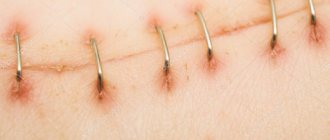
In general, a scar is formed in 4 stages:
- the first week and a half after skin damage. During this period, granulation tissue forms, blood vessels increase in size, swelling decreases and redness disappears. Proper care is especially important here - to prevent the wound from becoming infected, and also to avoid physical activity, which can cause the sutures to separate. If this stage is completed successfully, the wound will heal with careful primary intention.
- it continues for 20 days, starting from the 10th day after the injury. At this stage, the formation of the primary scar occurs. Its characteristic features are the formation of loose, easily stretchable tissue of a pink hue. This tissue is an external manifestation of the active production of collagen and elastin in the damaged area - fibers that are responsible for restoring the skin after damage.
- the next two months. A scar with a dense structure forms at the site of damage. Gradually, blood circulation in this area slows down, as a result of which it begins to turn pale.
- final formation, which lasts from 4 to 12 months.
As mentioned above, for plastic surgery to remove scars, a prerequisite is the formation of the scar. Firstly, because the final appearance of the scar can be quite aesthetic and satisfactory for the patient, even without the participation of plastic surgery. Secondly, until the end of formation, “fresh” tissues are prohibited from being “disturbed” by external influences, since this can negatively affect their further maturation.
Treatment options
There is a common belief that hypertrophic scars can be eliminated in various ways. This is wrong. With the help of some medical methods, you can only smooth out the scar, make it less noticeable and less rough. But it is impossible to completely eliminate a scar non-surgically.
Methods for treating hypertrophic scars:
- hormonal drugs (steroids);
- hyaluronidase;
- silicone bandages;
- Bucca Rays;
- cryodestruction;
- electrophoresis;
- laser therapy;
- mesotherapy;
- Microwave therapy;
- vacuum massage;
- near focus radiotherapy;
- therapeutic dermabrasion;
- Surgical dermabrasion.
Hormone therapy is an effective treatment for hypertrophic scars. Corticosteroids reduce inflammation, reduce the amount of connective tissue by reducing collagen synthesis and suppressing the activity of myofibroblasts.
Corticosteroids are given by injection and applied to the scar as ointments every day for two weeks.
But, as they say, every medal has a downside. You should be aware that hormone therapy can have negative consequences.
It can cause:
- thinning and sagging skin;
- formation of spider veins;
- the appearance of pigmentation.
Hyaluronidase is an enzyme capable of breaking down acidic mucopolysaccharides, including hyaluronic acid. This enzyme is used when there is a need to eliminate the effect of hyaluronic acid. For example, to eliminate excess hyaluronic acid when using fillers.
Hyaluronidase has a destructive effect on mucopolysaccharides, thereby helping fluids to move more actively under the tissues. As a result, swelling decreases, and the scar becomes smoother and softer. Hyaluronidase-based injections are injected directly under the scar or into the surrounding skin. To achieve the desired effect, from 6 to 15 injections are made daily. Usually the course is repeated after 2 months.
Silicone bandages must be worn around the clock for one year. But even wearing them gives a positive result only in 30% of cases.
Bucca rays are a method based on soft x-rays. Its essence lies in the fact that the hypertrophic scar is exposed to ionizing rays, which destroy myofibroblasts and collagen fibers. This method gives a good therapeutic result when scars have just formed.
However, this method has contraindications:
- age limit up to 16 years;
- kidney disease;
- skin diseases (dermatitis);
- diseases associated with circulatory disorders.
Cryodestruction has a good effect in the treatment of hypertrophic scars. The essence of the method is to administer liquid nitrogen under the scar or under the surrounding skin. The result of this manipulation is the crystallization of liquid inside and outside the cells, which leads to their death, and small vessels to destruction. After the procedure, a bubble forms at the site of the scar, which must be cut off and a special healing agent applied to the wound. This method is effective in 70% of cases.
Electrophoresis is used at the early stage of hypertrophic scar formation. Since it is at this stage that intensive synthesis of hyaluronic acid occurs. To reduce the scar and increase tissue permeability, electrophoresis is used with a specific enzyme hyaluronidase, which can reduce the viscosity of the intercellular matrix. As a rule, treatment consists of two 10-day courses with a two-week interval.
The treatment of an old hypertrophic scar will require three courses, but with collagenase (an enzyme that breaks down collagen).
Laser therapy helps reduce the volume of dilated vessels that cover the hypertrophic scar. The length and intensity of the laser beam depends on the condition of the scar tissue. The essence of the procedure is that scar tissue is removed layer by layer, followed by the regeneration of new cells. Laser therapy is analogous to electrophoresis, since both procedures give the same effect.
Mesotherapy involves puncturing the scar itself using hyaluronidase. The medicine is administered several times.
Microwave therapy is not an independent method of treating hypertrophic scars. As a rule, it is used in combination with cryodestruction. Microwave therapy promotes the transition of fluid inside the scar to a free state and is therefore easily eliminated by cryotherapy.
Vacuum massage causes a reflex effect by irritating skin receptors with the vacuum created in the flask. As a result, the circulation of lymphatic fluid and blood circulation improves. This method improves the condition of the skin, making it firm and elastic. Vacuum massage has a stimulating effect on scars, so it must be combined with dermabrasion.
Close-focus X-ray therapy affects fibroblasts with rays, making them less active. This method is especially successful in suppressing scar growth. But close-focus radiotherapy causes a number of complications in the form of skin depigmentation, malignant transformation of scar tissue and skin atrophy.
Therapeutic dermabrasion helps smooth out the skin and smooth out scar tissue. This happens in the following way: microcrystals falling on the surface of the skin “roll up” dead skin cells, gathering together in a special flask. The depth of exposure to the skin depends on the pressure in the vacuum. After the procedure, the skin is treated with special products. The number of procedures depends on the depth of resurfacing and the condition of the scar, as well as the reaction of the body itself.
Surgical dermabrasion gives a better effect than peelings, resurfacing and therapeutic dermabrasion combined, since it has a short treatment period. Its essence consists of layer-by-layer laser tissue removal and alignment of the scar with the surrounding tissue. After the procedure, it is necessary to treat this area of skin with a 2% boric acid solution.
Internal scars after mammoplasty
During breast augmentation surgery, after the doctor inserts the implant into the incision, he connects the internal tissues with threads from the intestines of small cattle. Cosmetic sutures are used to stitch the skin.
After the operation, they need to be treated, lubricated with creams that accelerate regeneration, to avoid tissue tension, a silicone patch is used. For 7 to 14 days after surgery, a woman must follow these rules:
- It is forbidden to strain your arms and upper body;
- You can't sunbathe;
- You cannot engage in active sports involving jumping or rhythmic movements for 2 to 3 months;
- It is contraindicated to raise your arms up, especially with minimal load;
- It is prohibited to drive a car;
- It is not recommended to have intimate relations, as this leads to increased blood pressure and increased heart rate.
In addition, the patient needs to wear compression garments.
If these rules are violated, the risk of divergence of internal seams increases. Then there is pain in the chest, swelling, asymmetry of the mammary glands is observed, and yellow liquid with a bloody admixture leaks from the wound.
If such symptoms appear, you must immediately consult a surgeon and perform an ultrasound. Depending on the degree of divergence of the internal seams, the doctor will recommend rest and shapewear or repeat surgery. It is impossible to solve such a problem on your own.
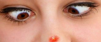
Keloid scars
Keloid scars are overgrown connective tissue at the site of a healed wound. This is an elastic, dense formation that protrudes beyond the boundaries of the wound and rises greatly above the surface of the skin. It has an irregular shape and visually resembles a tumor. Unlike other scars, keloid scars do not fade over time.
Keloid scars are considered a very serious cosmetic defect; they can cause limited mobility, as well as psychological discomfort.
The formation of keloid scars occurs in people of any age and gender, but is much less common in children and the elderly.
What influences scar formation? Factors.
- Age – skin heals more slowly as we age, while young skin tends to “over-heal”, older people develop larger, thicker scars than younger people;
- Genetic factors - skin type, people of African and Asian descent, with high skin pigment content, are especially prone to abnormal scars such as Keloids;
- Scar location - scars are largest near muscles that are particularly active, such as the back, legs, shoulders, near joints - they are often more noticeable than scars that form in less active areas;
- Wound infections or complications - an infected wound increases the likelihood of abnormal scarring.
Types of keloid scars
Keloid scars are:
- true;
- false.
A true keloid scar forms on its own and is more common in men in the chest area. Very often, such a scar can be mistaken for a tumor, which has a pink color, a shiny surface with visible veins of blood vessels. The keloid scar progresses rapidly, significantly increasing in size. The scar does not cause any pain and does not affect the general well-being of a person.
A false keloid scar forms after a skin injury: a burn, injury, postoperative wounds or a regular cut. Sometimes such a scar forms at the site of a healed ulcer, boil or abscess. When a keloid scar develops in place of an ordinary scar, its growth is initially unnoticeable. But gradually it grows into a huge bright pink tumor.
There is another classification of scars by age:
- young keloid scars, which range from several months to five years, are characterized by a bright color and a dense, smooth surface.
- old keloid scars, which are more than five years old, stabilized in growth, that is, they no longer grow, are characterized by a rough surface and pale color.
Clinical picture
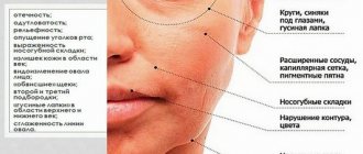
Symptoms of tympany in the early stages of development are manifested only by restlessness of the cow. She begins to refuse to eat food, fans her stomach with her tail, and constantly looks at her stomach because she feels bloated and uncomfortable. The animal begins to arch its back and moo, constantly trying to take a horizontal position, but immediately gets up and tries to kick itself in the stomach with its hind legs. Body temperature is normal, but breathing becomes much more frequent, it is chesty and shallow.
With the further development of the pathological process, the animal breathes through its mouth, often coughs, and groans. Bloating can accompany the development of other pathologies, so it is very important to visit a veterinarian as soon as possible, who will make a competent diagnosis and prescribe treatment.
Gradually, as the pathological process progresses, salivation increases, and foam is released from the mouth along with salivary fluid. Visually you can notice a swollen abdomen. The surfaces of the mucous membranes of the oral cavity become blue, the pulse becomes faster, and belching stops.
We invite you to familiarize yourself with the largest rat in the world: photos of a giant animal, what are domestic and outdoor animals called, the maximum size of Bosavi and bamboo rat
Causes of keloids
The reasons that contribute to the appearance of keloids are as follows:
- improper connection of the wound edges during surgery;
- infection and suppuration of the wound;
- malfunctions of the immune system;
- failure of the hormonal system;
- genetic predisposition.
However, the exact reasons for the formation of keloid tissue are still unknown, but it is known for sure that a keloid scar can form at the site of any wound due to disruption of the normal scarring process.
Introduction of feed after diet
A diet without chewing gum usually lasts 1-2 days. After this time, you need to start introducing regular food into your diet.
This must be done carefully so as not to provoke a relapse:
- On day 2-3 you can give some hay, silage or grass.
- Gradually add chopped vegetables, previously sprinkled with salt.
- It is useful to give mash made from grain flour with the addition of yeast.
- Unlimited water is required for the cow at any time.
- Gradually, regular food is introduced into the diet.
- If the reason for stopping chewing gum was improper feeding, it is necessary to reconsider the diet.
Childbirth is a great stress for any organism, regardless of whether it is a person or an animal. In cows, this condition can cause disruption of the chewing process.
When a calf is born, this happens for the following reasons:
- displacement of all internal organs in the cow’s body;
- psychological factors;
- postpartum trauma;
- mastitis.
To determine the exact cause, you need to call a veterinarian. Before his arrival, it is worth putting the animal on a food diet.
If sluggish chewing gum is observed, then drastic measures should not be taken. It is only important to adjust your diet. Knowledgeable people do not recommend using traditional methods for postpartum problems with the functioning of the stomach, because this can harm the cow.
We invite you to familiarize yourself with Raising a Scotch Terrier
If mastitis is a possible reason for the cessation of belching, all treatment actions should be aimed at curing it. You can massage the udder yourself. As the pain subsides, the belching reflex may return.
Providing the female with access to fresh air will help stabilize the functioning of the central nervous system and all organs.
Rumination is part of the cow's digestive process. The absence of chewing gum is a consequence of some disorder in the body. After eliminating the causes and introducing regular feed, the cow begins to live a normal life.
Many people wonder if a cow's temporary lack of cud has any consequences.
Important! In most cases, with proper and timely assistance to the animal, temporary cessation of chewing gum does not entail any negative consequences.
The most dangerous moment is when a pregnant cow, due to compression of the diaphragm by the fetus and a full stomach, suffocates. If you do not pay attention to the cow’s condition in a timely manner and do not resort to help, the animal will simply suffocate from lack of air. The owner will suffer large losses from the loss of the individual.
Also, if a foreign body enters the digestive organ, the walls of the gastrointestinal tract may be injured. And removing an object from an animal’s body is also not an easy procedure. Usually minor wounds heal on their own, and external visible damage is treated.
Maintaining cleanliness in the barn will ensure rapid healing of post-operative sutures.
Sometimes there is a decrease in the weight of cows and a decrease in the level of milk production in the absence of the ruminant process. But this phenomenon is quite rare, since people have learned to cope with this illness.
The gastrointestinal tract of a cow is designed in such a way that food must travel a long and long way to be completely absorbed. The ruminant process is normal for cattle. Any disruption in the functioning of the natural systems of the body requires immediate action to eliminate the causes and the provision of all possible actions to save livestock. Otherwise, financial losses will significantly hit the owner’s pocket.
Symptoms of keloid scars
As a rule, the favorite location for keloid scars is the chest, back, and shoulders, but this does not mean that a keloid cannot appear in another anatomical area.
In the process of formation, keloid scars manifest themselves as follows:
- first there is a noticeable cosmetic defect;
- as the scars develop, they itch, burning and tingling occur;
- painful sensations are felt when pressing on the scar;
- there are some restrictions in the mobility of the limbs;
- When the skin in the scar area is damaged, the healing process takes a very long time.
Causes of the disease
Preventative measures are always the best option rather than treating the animal.
To protect the cow as much as possible from the condition associated with the lack of cud, it is worth taking the following measures:
- monitor the cleanliness of feed and water;
- do not allow small objects to get into the feed;
- do not overload your diet with hard foods;
- grind feed intended for young animals;
- feed livestock regularly, following the regime;
- maintain cleanliness of the premises;
- calves can sometimes be given mineral water to stimulate the stomach;
- provide the animal with sufficient water;
- monitor the general condition of the livestock.
As for the lack of cud in a pregnant cow, it is important to add gentle nutrition to the above measures and not overfeed too much.
Why doesn't the animal burp? Today, veterinary medicine divides tympany into two forms: primary and secondary. And also for subacute, acute and chronic course of the disease.
An acute disease of the primary stage occurs due to eating large quantities of beets and potatoes, ripe clover or alfalfa, legumes, wet grass, young crops and rotten apples. The development of the disease occurs at a faster rate if the cow is sent to water immediately after eating the specified food.
The second form of the disease occurs due to the consumption of poisonous plants (monkshood, hemlock, colchicum, poisonous plant). At this time, tympany occurs after rapid paralysis in the walls of the scar. Also, the stomach may swell during disturbances in processes such as belching and chewing gum, or due to blockage of the esophagus itself.
The course of the chronic form of the disease continues for a long time, and the development of the disease is facilitated by chronic gastritis and the traumatic form of radiculitis.
The acute form of the disease requires immediate treatment. During chronic deficiency, it is enough that you simply get rid of the causes. If this is ignored, the animal will quickly lose weight, gradually lose milk production, and ultimately the farmer will have to send the sick cow to slaughter.
The first signs of the presence of the disease can be detected by observing the general behavior of cattle. From the very beginning, the cow behaves restlessly and refuses to eat food. At this time, the animal begins to experience unpleasant and, in some cases, painful sensations in the anterior abdomen and chest.
- Breathing becomes heavy, shallow and rapid (60-80 breaths per minute), there is a cough;
- The veins on the cow's head and eyes are increased in size;
- The mucous membranes acquire a blue tint;
- The pulse reaches 100 beats per minute;
- Foamy saliva comes out of the mouth;
- Initial strengthening, and then complete cessation of scar movement;
- Vomiting;
- The hungry pit is leveled out and the volume of the abdomen becomes noticeably larger.
If the farmer does not provide assistance to the animal in the first 2-3 hours of the disease, it will not survive. During acute periodic bloating, the animal is much more anxious than during foamy bloating. In this case, all the above symptoms of the disease are noted.
The chronic form of the disease is characterized by milder symptoms, which are observed after eating food. At this time, there is a gradual decrease in the total weight of the animal. The acute form of the disease in the absence of timely and comprehensive treatment can lead to the death of the animal.
The duration of the chronic form of the disease ranges from one week to several months. If active treatment is not started during this time, the animal will also die.
Stages of development of keloids
A keloid scar has several stages of formation:
- epithelialization - the damaged area of the skin becomes overgrown with a layer of epithelial cells, which become rough and dense;
- swelling - occurs during the growth of the scar, causing unpleasant painful sensations;
- compaction is almost the final stage of keloid formation, so the scar acquires its characteristic appearance;
- softening – at the very end of scar formation, it becomes soft, mobile and painless.
Treatment of keloid scars
Since there are no generally accepted methods for treating keloids, symptomatic therapy is carried out, which is aimed at eliminating unpleasant symptoms. Thus, treatment is selected individually in each specific case:
- the use of ointments and creams based on hormonal drugs can be effective in the treatment of young keloids that have just formed;
- laser resurfacing of the scar, which gives good results in smoothing the keloid, but does not lead to its complete disappearance;
- cryotherapy is effective only in the treatment of young scars, since old keloids do not respond to exposure to low temperatures;
- Surgical treatment is carried out only in cases of large keloid scars, but after excision of the scar, a new one may form in its place, especially if the patient has a hereditary predisposition to them.
Plastic surgery scar removal
It was said above that the choice of scar removal method is influenced by several factors, among which, in addition to the type, its localization is also important. So, for example, if the scar is located on the face, the surgeon will suggest a technique for cutting out the scar, followed by stitching the edges of the wound using an internal suture technique, located not on the surface of the skin, but inside - parallel to it, and using the thinnest threads, less than a human hair in diameter. If the scar is located on another part of the body, then it may be appropriate to carry out laser excision there, or resort to dermabrasion.
In addition, the decisive factors
here will also become:
- the location of the scar relative to the natural tension of the skin;
- its size (affects the nature of scarring, large-scale injuries are healed more “reliably” and less aesthetically pleasing than small and subtle injuries);
- the age of the patient (in children and adolescents, wounds heal quite quickly, in people over 30, regeneration processes in the skin proceed much more slowly, and prolonged healing can negatively affect the results of plastic surgery for scar removal);
- genetic predisposition to unaesthetic healing of wounds and injuries. For the surgeon, the condition of the scar can become a guideline for choosing a scar removal technique. If the patient is predisposed to the formation of rough scars, the surgeon is more likely to suggest resurfacing.
How are hypertrophic scars different from keloids?
After healing, a flat scar forms at the site of any wound. This is a normal process of tissue scarring. When the deeper layers of the skin are damaged, rough and dense scars appear, since a large amount of connective tissue is used for their healing. But in some cases, the connective tissue grows excessively, forming hypertrophic and keloid scars.
A hypertrophic scar is always limited by the size of the wound, that is, it never extends beyond its boundaries. Sometimes hypertrophic scars can disappear with steroid treatment. This process can continue for more than a year.
Keloid scars may not form immediately after injury, but after some time. They always go beyond the boundaries of the wound, growing more and more. The ability to overgrow and invade adjacent healthy tissue distinguishes keloids from hypertrophic scars. In addition, they appear not only after an injury, but also as a result of mild inflammation of the skin, for example, a slight burn, a pimple, or a small papule that was not even damaged.
Traditional ways of providing assistance
The absence of cud in a cow indicates problems with digestion. Often the disease can be triggered by very serious reasons. There are situations when there is no time to wait for the veterinarian to arrive - the animal needs to be saved, otherwise it may die. Therefore, you need to be prepared for any possible situation. A person is capable of providing first aid until a specialist arrives.
People started raising cows a very long time ago. In those days there were no veterinarians or medicines, but since livestock was the main source of food, it was protected and valued. Therefore, people unwittingly learned to help animals with the help of improvised means and scavenged materials.
To this day, traditional methods of treating cows are popular among breeders, especially on personal lands with a small number of livestock. The recipes have been proven over time and there is no doubt about their effectiveness.
- Starvation diet for a cow. She only needs to be given plenty of water throughout the day.
- Hellebore tincture stimulates the digestive system. Every 30 minutes, the cow is given half a liter of water with 10-15 ml of hellebore.
- Add tinctures of wormwood, St. John's wort and chamomile to your drink. An effective bloating reliever.
- Stimulation of organ function with mineral hydrochloride water (approximately 2 liters).
- Cucumber pickle (sometimes cabbage or tomato pickle) is also good if the cow does not have cud. 0.5 liters 3 times a day.
- Sometimes a cow is given a few liters of freshly milked milk to drink to get the gastrointestinal tract working.
- You can drink 150 ml of vodka, which normalizes the functioning of the stomach. The main thing is not to overdo it, so as not to cause poisoning.
- Acetic essence in an amount of 40 ml, diluted in 2 liters of water with the addition of 500 grams of sugar, restores protein functions in the body.
- Sometimes, in the absence of belching, vodka with garlic is used. 2 tablets of no-shpa and a little vegetable oil are placed in the cow's mouth. After this, pour the tincture of vodka and garlic into the throat in the proportion of 2 cloves per 0.2 liter of liquid. The functioning of the gastrointestinal tract is restored in approximately 4-5 hours.
- To start a cow's stomach, you can prepare the following solution: 50 ml of alcohol, 100 grams of live yeast, 0.2 kg of granulated sugar and 1 liter of water.
Traditional methods of treatment must be used carefully. You can't give a cow everything at once. You need to choose the most optimal treatment. If it doesn't help, then you need to try something else. Some time should pass between doses (at least an hour).
Of the tinctures containing alcohol, you need to choose only one, since an excess of alcohol will cause poisoning of the body.
During the period of illness, it is important to carefully monitor the sick individual. If the folk remedies used do not bring the desired effect, and the animal still does not regurgitate food for chewing, you need to seek the help of a specialist. The cow may need surgery if the cause of the lack of cud is a foreign object in the digestive tract.
Scar Prevention
It is difficult to avoid the formation of a keloid scar, especially when it appears on the site of a completely harmless pimple, but you should still follow certain tactics after any damage to the skin:
- Immediately consult a doctor to treat the wound and apply sutures (if necessary);
- wear special compression garments that inhibit the growth of connective tissue;
- use special ointments that suppress the growth of scar tissue;
- avoid exposure of the wound to ultraviolet rays;
- avoid any mechanical impacts in the scar area.
To summarize, it is worth remembering once and for all that all methods of treating hypertrophic scars and keloids can only smooth out their appearance against the background of healthy tissue, make them less pronounced and pale, but it is impossible to completely eliminate scars.
Features of postoperative internal scars
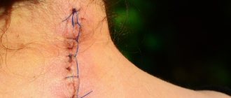
Surgical sutures can be internal or external. To apply the former, threads from the intestines of small cattle are used, thanks to which the tissues are firmly connected. External sutures temporarily secure the edges of the wound so that the scar can form faster. They are made with threads made of linen, silk or synthetic material.
External sutures are removed after 6 to 14 days, and internal sutures usually dissolve on their own.
If the patient does not follow the rules of behavior after surgery, for example, lifting weights or playing sports, then the internal scars after surgery may disperse. This is evidenced by:
- Painful sensations;
- Increased body temperature;
- Slowing heart rate;
- Dizziness.
If such symptoms occur, you should immediately visit a doctor.
The specialist will examine the wound and prescribe the necessary tests (blood test, ultrasound) that will help determine the degree of its discrepancy, the location of the accumulation of ichor or pus. Treatment consists of limiting physical activity, taking antibiotics, physical therapy, etc.
If the discrepancy of the internal scar intensifies, then surgical intervention cannot be avoided. In case of severe concomitant pathologies, for example, diabetes mellitus, a temporary or permanent mesh is applied to the damaged area.