Serous mastitis is a disease of the mammary gland caused by pathogenic bacteria. The causative agent in most cases is Staphylococcus aureus, a microorganism that is very resistant to antibacterial drugs. Most often, mastitis occurs in women during breastfeeding in the first months after childbirth. Bacteria enter the mammary gland through cracks, wounds and lymphatic vessels, causing inflammation.
There are 2 types of serous mastitis: lactational and non-lactational. Cases of the non-lactational type account for only about 5% of the total number of mastitis. A disease not associated with feeding, as a rule, proceeds less violently, but can become chronic with periodic relapses.
Causes of serous mastitis
In the serous form of mastitis, the parenchyma and milk ducts of the gland become inflamed. The infiltrate does not consist of pus, but of lymph and intercellular fluid.
Causes of the disease:
Incorrect hygienic breast care during breastfeeding - rubbing the nipple, infrequent or too frequent wiping or washing of the breast.
Installation of an artificial implant, compression with synthetic material, disruption of lymph outflow.
Incorrect grip of the nipple during feeding: the baby pulls it without covering part of the areola with his mouth.
Stagnation of milk and disruption of its normal outflow.
Stagnation of milk, or lactostasis, leads to impaired absorption and secretion of lactic acids. As soon as the serous fluid penetrates the breast tissue, the outflow of lymph becomes difficult and an infiltrate forms. The appearance of a serous infiltrate leads to inflammation of the gland tissue. If infectious microorganisms penetrate into the breast through damage in the form of cracked nipples, serous mastitis transitions into a purulent form.
Reasons for the development of lymphostasis and swelling of the hand
The main prerequisite for the transition of soft edema of the hand to the middle stage and to dense edema is improper rehabilitation. If during the early postoperative period conditions are not created for the outflow of lymph from the arm area, complications are inevitable.
Fluid stagnation is caused by:
- mechanical obstacles to the movement of lymph (clamping of the hand, adhesions as a result of irradiation of the tumor);
- injuries to the problematic arm (even minor ones, for example, from a blood pressure cuff or a needle prick) - hematomas in places of lymphostasis take a long time to resolve and not always completely, sometimes a benign tumor forms at this site, which creates a mechanical obstacle and can lead to necrosis;
- excessive physical activity - in this case, muscle tissue develops on the arm, the amount of protein increases, which means an increased amount of fluid is attracted;
- neglect of rehabilitation procedures (physical therapy, massage, medication);
- wrong lifestyle.
Diagnosis of serous mastitis
In order not to give up breastfeeding, you need to detect signs of the disease in time. The doctor should know exactly when breast problems began. The doctor should monitor changes in body temperature. If it is elevated, general antibacterial therapy is carried out using broad-spectrum antibiotics.
On palpation, a painful lump on the chest is noted. In some cases, clear liquid is released from the nipple even with slight pressure on it.
Another diagnostic method is ultrasound. With this research method, it is possible to accurately determine the location of the infiltrate or the presence of lactostasis.
Why does the nipple discharge clear fluid?
Many representatives of the fair sex notice clear discharge from their nipples. But this does not always mean the occurrence of some pathology. The fact is that, due to the physiological characteristics of women, the appearance of such transparent fluid from the mammary gland is considered normal, especially during pregnancy and breastfeeding. However, to identify the exact cause, it is best to consult a mammologist for advice. It is worth noting that sudden discharge from the breasts of a non-breastfeeding woman is a rather alarming sign of the development of various serious diseases. Therefore, it is very important to visit a specialist to conduct a series of studies, based on the results of which the existing pathology will be identified and appropriate treatment therapy will be prescribed.
As a rule, a comprehensive examination includes: blood tests, MRI, ultrasound of the mammary glands or mammography, cytological examination of secretions, ductogram with the introduction of a contrast agent. The most common causes leading to discharge from the mammary glands include: dilation of the milk ducts, mastopathy, galactorrhea, acute or chronic diseases of the pelvic organs, condition after an abortion or spontaneous miscarriage, purulent diseases of the mammary gland, closed breast injury, mastitis, breast cancer glands, intraductal papilloma, and Paget's disease.
The main questions that the doctor will definitely ask during the consultation
As a rule, during an appointment, the mammologist necessarily asks the patient the necessary questions that will help establish the most accurate diagnosis: what color of discharge comes from the nipple, fluid is released from one mammary gland or from two, how often does this happen, whether this discharge comes out of the nipple on its own or immediately after pressing it, has there been a chest injury, are there any other disturbing diseases with elevated body temperature, accompanied by headaches, malaise and blurred vision, is there any course of treatment with any medications?
Treatment and preventive measures
It must be remembered that constant discharge from the mammary gland should never be ignored.
Basically, therapeutic therapy includes the use of hormonal drugs, traditional methods of treatment, and antibiotics. In addition, in exceptional situations, surgical intervention is recommended. Experts advise women who find themselves with nipple discharge to strictly observe hygiene of the chest area. The mammary glands should be washed in the shower at least twice a day, and then dried thoroughly. It is also advisable to wear a soft bra that does not compress the breasts. Why does a clear liquid come out of the nipple www.kakprosto.ru
Mastitis in a nursing mother
In relation to the total number of births, the frequency of mastitis ranges from 3 to 20%. In most cases, purulent inflammation in the mammary gland in women in the postpartum period is caused by S. aureus (Staphylococcus aureus). The “entry gates” for pathogenic microbes are cracks and microtraumas of the nipples. It is possible that infection can enter through the milk ducts during feeding or pumping. Mastitis in a nursing mother can be a consequence of improper care of the mammary glands or develop due to non-compliance with personal hygiene rules.
Postpartum mastitis, unlike its other forms, is predominantly associated with lactation (hence the name “lactation”) and is diagnosed in 2-11% of lactating women. Lactation mastitis is characterized by unilateral damage to the mammary glands, develops mainly at 5-6 weeks after birth and goes through all the stages inherent in acute purulent mastitis of any origin.
Violation of the regimen and/or rules of breastfeeding provokes stagnation of milk in the mammary gland, which provokes the development of local non-infectious inflammation.
Since the trigger for the development of inflammation is lactostasis, at the beginning of the disease a woman experiences a feeling of tension in the mammary gland. Stagnation of milk leads to the fact that the mammary gland increases in size, and overcrowded milk ducts can be felt in the form of painful compactions without clear boundaries. The amount of expressed milk decreases significantly and body temperature rises.
If lactostasis is not eliminated in the next 3 to 4 days, secondary pathogenic flora attaches, which causes decomposition of milk and damage to the milk ducts, that is, the process takes on the character of acute purulent inflammation. The mammary gland looks swollen and red, the discharge from the nipple becomes purulent, and symptoms of intoxication increase. An attempt to empty the mammary gland is not possible due to severe pain. The further scenario of the disease depends on how quickly the patient seeks qualified help: if a woman does not turn to a specialist, does it too late, or tries to cope with the disease on her own, the likelihood of developing a severe infectious process becomes very high.
Non-lactation mastitis after childbirth is much less common; it develops without the participation of lactation and is similar to that in non-lactating women.
Preventive measures for the development of mastitis
Preventive measures to prevent serous mastitis are carried out during pregnancy and after childbirth. In the prenatal period, prevention consists of measures aimed at increasing the pregnant woman’s body’s resistance to infectious diseases, namely, identifying and timely eliminating various complications. This also includes the rehabilitation of foci of chronic inflammation: caries, inflammation of the urinary system, etc.
For flat or “inverted” nipples, doctors recommend performing a special mass, which can be easily done at home. However, keep in mind that the effect on the nipples can lead to contractions of the uterus, so the effect should be light and short-lived.
Basic preventive measures in the postpartum period:
- mandatory observance of personal hygiene rules;
- softening the breast skin to avoid cracks;
- prevention of lactostasis (milk stagnation);
- feeding on demand, without time limit;
- expressing leftover milk after feeding;
- wearing loose bras and special nipple covers.
Risk factors for serous mastitis also include nervous tension and stress. Therefore, it is so important to monitor psychological balance, maintain a daily routine, get good sleep and eat well. Prevention of mastitis not associated with lactation lies, first of all, in observing hygiene rules and timely diagnosis and treatment of skin diseases.
Many women, when symptoms of mastitis appear, do not rush to see a doctor and use traditional methods of treatment: herbs, compresses, massage. Experts do not recommend treating mastitis at home. The insidiousness of this disease lies in the difficulty of determining its form and stage, and folk remedies do not have the necessary effectiveness to completely destroy the infection.
Improper use of herbs and other remedies can lead to the development of purulent forms of mastitis, which poses a danger not only to the health, but also to the life of a woman. Traditional methods are recommended only as an auxiliary therapy and only after the approval of the attending physician.
Classification of mastitis
Mastitis is classified as follows:
With the flow:
Acute - characterized by all the clinical manifestations of mastitis, with severe chest pain.
Chronic – characterized by a weakening of the clinical manifestations of the disease with periodic exacerbations. Upon palpation, a dense, inactive seal fused to the skin is determined.
Forms of mastitis
Lactation mastitis (postpartum) is primarily a consequence of prolonged lactostasis during breastfeeding. It is characterized by breast swelling, sharp pain when feeding or expressing milk, redness of the skin and a rise in body temperature. The milk may contain impurities of pus and blood.
Plasma cell mastitis is a rare type of disease caused by multiple births. It develops mainly after lactation and in women after 30-40 years. It is characterized by processes of infiltration of tissues under the nipple with plasma cells, and is also accompanied by processes of hyperplasia of the excretory mammary ducts. There is usually no formation of pus. Clinical manifestations may resemble breast cancer.
Mastitis of newborns - just like in adults, it is characterized by swelling of the mammary glands with discharge when pressed, only not only in girls, but also in boys. Inflammation is caused by the residual effect of sex hormones transmitted from the mother. Clinical manifestations usually subside after a few days, however, in some cases medical attention may be required.
By the nature of inflammation
- Serous is the initial stage of the development of pathology, during which serous fluid appears in the mammary gland.
- Infiltrative is the next stage of the development of the disease, in which serous fluid from the mammary gland seeps into the external tissues, forming foci of infiltration, while the breasts become full and the skin becomes stretched.
- Purulent is the third stage of mastitis development, in which infiltrates are transformed into purulent deposits.
- Abscess – characterized by a limited purulent focus in one place, localized.
- Phlegmonous - characterized by widespread purulent formation in the chest.
- Gangrenous – characterized by the formation of necrotic foci during a long course of a purulent inflammatory process.
Types of discharge from the breast with mastopathy
It is common for every woman to take care of her health. The chest is an area of increased attention. Having noticed even small changes, most people rush to see a doctor. One of the most common complaints when visiting a mammologist is the appearance of discharge from the mammary glands.
Breast discharge often accompanies mastopathy
It should be remembered that during some periods of life, such as pregnancy or subsequent lactation, discharge from the breast is considered normal. The appearance of fluid from the nipples immediately after stopping breastfeeding and a few days before the onset of menstruation should not cause concern. In all other cases, a woman should undergo an examination to find out the cause of the discharge. According to experts, the main pathology with these symptoms is mastopathy.
Mechanism of discharge formation
The mammary glands are designed for lactation. This function determines some features of their structure. The main tissue of the female breast is glandular. It is penetrated by numerous water ducts, which merge into the mammary cavity and open outward in the form of pores at the top of the nipple. During pregnancy, colostrum production begins in the depths of the mammary glands, which is then replaced by milk.
With a pathology such as mastopathy, under the influence of hormonal imbalance, glandular tissue is destroyed and connective tissue grows. The work of the secretory mechanism is restructured, the contents of the water ducts change, and the woman notices uncharacteristic discharge from the nipples.
Mastopathy is characterized by an increase in connective tissue in the mammary glands
Character of discharge from the chest
Discharge from the nipples with mastopathy is an optional sign. In some cases, the disease proceeds without them. The appearance of fluid from the chest may be the first sign indicating incipient problems in the body, or it may signal serious pathologies.
The fluid secreted from the breast is of a serous or colostrum nature; most often it is transparent, without impurities, but with a characteristic odor. The discharge can be either copious, leaving marks on the underwear, or scanty, appearing only with strong compression of the nipple.
Since mastopathy is characterized by parallel development in both mammary glands, fluid is usually released from each nipple. The process of fluid secretion is often accompanied by painful sensations.
Women usually notice discharge of the following colors:
- colorless;
- white;
- yellowish;
- brown;
- green;
- red.
The color of the discharge may depend on the stage of the disease, its shape, and the size of the tumors. So, at the very beginning of the development of mastopathy, the discharge may be absent or transparent. A woman is concerned about the secretion of fluid from the mammary glands not on any day of the cycle, but only in the second half. After menstruation, the discharge from the breasts stops. This process is controlled at the hormonal level. As the cysts grow, the color of the secreted fluid changes to a more intense color. When the lumps reach a large size, a yellow or yellow-brown secretion is released from the nipples.
The green color of the discharge is due to the admixture of pus produced when an infection develops in a cyst or node. The bloody nature indicates serious complications, from intraductal papilloma to breast cancer.
The appearance of any discharge is a reason to undergo an emergency examination.
Nipple discharge may vary in color depending on the stage of the disease.
Discharge from the mammary glands in different types of mastopathy
A pathology such as mastopathy can exist in two forms: diffuse and nodular. The nature of nipple discharge often differs in different forms.
In the diffuse form, when there is an uneven growth of connective tissue, the fluid released from the breast is similar to colostrum. It is often clear or whitish, and its volume varies depending on the phase of the menstrual cycle. Greenish or light yellow discharge from the mammary glands occurs rarely, in approximately 5% of affected women.
One of the most common types of diffuse form is fibrocystic mastopathy. It is almost always accompanied by discharge due to deformation of the glandular tissue by overgrown cysts. The whitish character of the secreted fluid can be due to impurities of separated epithelial cells. When the capillaries passing through the tissue are damaged, a small amount of blood appears in the secretions.
The nodular form of mastopathy is accompanied by yellowish or greenish transparent discharge from the nipples, appearing a few days before the onset of menstruation. They can appear when pressure is applied to the nipple, but in some cases spontaneous secretion also occurs. The green tint is due to prolonged stagnation of secretions in the ducts, which are compressed by the nodes. In rare cases, with nodular mastopathy, bloody discharge from the mammary glands occurs. This is a complicated course of the disease that requires surgical intervention.
Nodular mastopathy may be accompanied by transparent yellowish-green discharge
Mastopathy or cancer. How to distinguish?
If the discharge has acquired a brownish or dark yellow tint, and its smell has intensified, then this indicates an advanced stage of mastopathy. It is known that if left unattended, this disease can degenerate into a malignant tumor. Such formations are developing rapidly, so it is extremely important to respond to changes in a timely manner.
The appearance of even a slight admixture of blood in the fluid released from the nipples indicates a possible cancerous tumor. In this case, it is worth undergoing diagnostic surgery.
What to do if you notice discharge
If pathological discharge appears from the nipples and mastopathy is suspected, the woman should visit a mammologist and undergo a series of additional examinations. Based on the data obtained, the specialist will determine a treatment regimen for mastopathy and rule out the presence of more serious pathologies.
As a rule, a woman is recommended to undergo the following examinations:
- ultrasonography;
- computed tomography;
- magnetic resonance imaging;
- mammography.
Additionally, a number of laboratory tests may be required, such as determining the level of hormones in the blood, cytological examination of fluid secreted from the mammary glands, and analysis for genetic predisposition to the development of neoplasms.
Mammography helps identify the causes of nipple discharge
Treatment of breast discharge
Secretion of fluid by the mammary glands is one of the symptoms of mastopathy, so getting rid of this symptom is directly related to the treatment of the disease itself. The doctor prescribes complex therapy, including taking hormonal medications and vitamins.
Until the discharge from the mammary glands stops, a woman must maintain careful hygiene. Daily procedures should include washing your breasts with warm water and changing your bra.
You should stop wearing underwear made of synthetic fabrics. For large volumes of discharge, it is permissible to use special hygienic liners.
Taking care of breast health is an important aspect of a woman’s life. In most cases, nipple discharge is not normal. A similar phenomenon in mastopathy requires increased attention. Regular self-examinations, medical examinations and consultations with a mammologist will help avoid serious complications.
vrachlady.ru
Signs of illness
Often the disease develops as a result of congestion in the mammary glands. The symptoms of serous mastitis are similar to lactostasis:
These diseases can be distinguished by the location of the increase in body temperature. With lactostasis, the temperature rises mainly in the armpits, since this is where stagnation develops. Thus, the temperature in these areas will always be higher than in the zone where there is no stagnation.
With serous mastitis, the temperature also rises, but the difference in its values in each armpit is insignificant. Another distinctive feature is that with serous mastitis, the body temperature does not fall, even after the glands are emptied, as with lactostasis. The woman's general condition is also not getting better.
Preventive actions
In order to avoid mastitis in dairy herds, it is necessary to strictly observe all the rules for keeping animals and raising young animals. It is imperative to treat the udder before and after milking. Use disposable wipes to wipe your nipples.
Monitor the quality of feed and the completeness of the diet. Conduct conversations with machine milking operators, farmers and private owners keeping cows.
It is necessary to conduct tests at least once a month for the subclinical form of the disease. Prevention of cow mastitis helps to avoid large time and financial costs for treatment.
Folk remedies for mastitis for a nursing mother
Treatment of mastitis in a nursing mother at home with fever must be previously agreed with the attending physician, otherwise the patient's condition may worsen, which will lead to the development of complications. In most cases, a course of antibiotics is indicated to treat the disease.
When treating mastitis at home, it is important to follow several rules:
- continue to express breast milk from both breasts;
- you should not use medications to reduce lactation;
- do not take hot showers or baths;
- do not heat areas of inflammation;
- Do not prescribe antibiotics yourself.
Dill seeds
Dill seeds have a rich biochemical composition, which has a pronounced antispasmodic and bactericidal effect, thereby reducing pain and inflammation. Compositions based on dill seeds have a diaphoretic effect, which reduces tissue swelling and temperature.
Recipe No. 1:
- 1 tbsp. dill seeds pour 50 ml of chilled boiled or purified water;
- put on low heat and simmer for 4-5 minutes;
- Take small sips throughout the day.
Recipe No. 2:
- Pour 100 g of dill seeds into 500 ml of milk and boil the infusion for 20 minutes;
- leave for 2 hours;
- Take a glass three times a day before meals.
Cabbage leaf
Compresses for mastitis in nursing mothers made from cabbage leaves relieve inflammation and stimulate blood circulation. To provide a pronounced therapeutic effect, it is recommended to use homemade cabbage. The leaves and the middle of the head of cabbage are used.
Recipe No. 1:
- Before applying a cabbage leaf to an inflamed chest, it is recommended to beat it well from the inside with a wooden hammer. This is necessary for the active release of juice.
- The inner side of the sheet is applied to the mammary gland and securely fixed with an elastic bandage, adhesive plaster or other bandage.
- You need to keep this compress throughout the night.
It is recommended to repeat the procedures until complete recovery.
Recipe No. 2:
- Grind the cabbage leaf and add a small amount of yogurt to it.
- Transfer the resulting composition onto a piece of cotton rag and apply to the inflamed mammary gland.
- The compress can be worn constantly, replaced with a new one once within 2-3 days.
Natural honey
Honey is a universal folk remedy that is used to treat many diseases. Its effectiveness is due to its rich biochemical composition.
Recipe No. 1:
- To obtain the medicinal composition, you need to mix natural honey, aloe juice and corn oil in equal proportions.
- Mix all ingredients thoroughly and refrigerate for 12 hours.
- Remove the product from the refrigerator and allow it to warm up naturally.
- Apply a small amount onto a cotton cloth and apply to your chest.
Recipe No. 2:
- Bake a large onion in the oven.
- Mix the juice released from the onion with honey.
- The composition is used in the form of a compress.
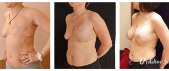
First aid and treatment
The main rule is not new: it is easier to prevent dangerous complications of breast removal than to get rid of them. Therefore, it is recommended to begin restoring arm mobility as early as possible after surgery.
Important! Physical activity should become daily, but overload is unacceptable: you cannot lift or carry weights of more than 1 kg; prolonged efforts require periodic rest.
Main activities within the framework of rehabilitation:
- drug prevention of infectious complications - carried out in the hospital before removal of the mammary gland, during and after surgery;
- bandage the limb (neither tight nor weak) with an elastic bandage to avoid tissue stretching;
- physiotherapy - also prescribed in a hospital, then adjusted by the attending physician - hardware maintenance of the elasticity of hand tissues and elimination of swelling - procedures using magnetic and ultrasonic waves, without heating;
- diuretics – prescribed by a doctor as a short-term measure, once or periodically;
- manual therapy - lymphatic drainage massage of the hand, which the patient learns to do independently over time, alternates with a general body massage performed by a specialist;
- physical therapy - a special set of exercises to gradually develop the muscles of the arm and increase the elasticity of the ligaments in the area of the operation, which is taught in the hospital - is performed 2 - 3 times a day during the first year, as well as swimming several times a week.
Prevention
Preventive measures will help avoid the development of mastitis. The quality of care for the cow and the conditions under which it is kept are of great importance.
It is also important what her diet is. Balanced food containing all the necessary vitamins and substances will help strengthen the animal’s immune system
What needs to be done to prevent serous mastitis in cows:
- Maintain cleanliness in the area where animals are kept.
- Observe the rules of udder hygiene.
- Handle this part of the cow's body with care - inspect for damage, wash, and milk carefully.
- Express the udder regularly and until completely empty.
- Do a breast massage.
Serous mastitis is very dangerous - the disease not only reduces milk yield and makes milk unsuitable for food, but also threatens the life of the animal. If the first symptoms of illness are ignored, the cow may begin to experience tissue necrosis, which often leads to sepsis. Careful attention to livestock and prevention of mastitis will help avoid the development of the disease.
Beautiful breasts are the adornment of every representative of the fair sex. However, there are a number of diseases that can cause breast problems. One of them is mastitis. This is the name for inflammation of the breast tissue. Many women do not pay due attention to the first signs of the disease and seek help when serous mastitis (the initial form of the disease) progresses to more severe stages. Acute mastitis is usually diagnosed in young nursing mothers and can occur in purulent and non-purulent forms.
Classification and stages of development
Initially, serous fluid consists of a small number of leukocytes and protein. When the disease begins to actively develop, white blood cells begin to die, as a result of which the liquid becomes thick and has a yellow tint. The process of putrefaction begins in it, which spreads into nearby healthy structures - infiltration. This phenomenon gives the name to the stage of the disease - infiltrative, and its form - acute infiltrative mastitis.
The structure of the mammary glands is considered to be a factor that contributes to the infiltration process. There are no dense connecting partitions between the individual parts of the tissues that prevent the active development of purulent mastitis. When suppuration spreads into healthy tissue, the disease enters the stage of acute destructive mastitis.
A purulent inflammatory process in the mammary glands can lead to the appearance of abscesses (an abscess that is located inside the capsule) or phlegmon (the abscess is not limited to the capsule from tissues that are not yet affected), which can be located near the nipples, under the skin, behind the mammary gland.
Purulent or acute destructive mastitis is more difficult to resolve than serous and infiltrative mastitis. It is necessary to remove the accumulated pus through surgery so that the disease does not affect healthy tissue outside the mammary gland and does not lead to sepsis.
Fluid in a breast cyst: is it dangerous?
Liquid in the tumor is not considered a dangerous pathology. This is absolutely normal for a cyst. Its contents may have a transparent or yellowish tint. The danger comes from a cyst that does not cause any symptoms for a long period of time.
Because an inflammatory process may develop. It is characterized by suppuration in the cyst, and the capsule is filled with a thick liquid - pus. In such situations, cancer cells may appear.
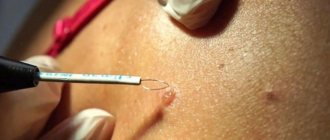
Therefore, for diagnosis it is necessary to undergo a special analysis - a biopsy. It is a small puncture in the cyst to examine the fluid. Based on the result of this analysis, the attending physician determines the required treatment.
In some cases, the cyst does not need to be treated; it can simply be observed. Over time, it dissolves on its own. More information about whether a breast cyst can resolve and what contributes to this can be found here.
Symptoms of serous mastitis
The symptoms of the disease are quite common at first, and resemble a common cold, but after 2-3 days, mastitis becomes acute and its symptoms become quite specific.
- Increase in body temperature, sudden jumps to 39-40 degrees, followed by a decrease to normal temperature.
- Chills, fever and other common symptoms of inflammation.
- Redness of the skin at the site of inflammation.
- Poor milk flow, sometimes inability to continue feeding.
- Bursting pain in the mammary gland.
- Swelling of the gland, redness of the skin at the site of inflammation.
Serous mastitis begins like a regular flu; most women, experiencing malaise, weakness, and drowsiness, attribute the symptoms to a cold. A little later, specific symptoms are added to the familiar ones: chest pain, redness of the skin at the site of inflammation. Body temperature rises, breasts swell and feeding becomes difficult
It is important to recognize mastitis immediately and consult a doctor immediately. Signs of acute mastitis require immediate treatment
Redness of the skin is an external sign of an inflammatory process in the chest
Diagnostic methods
In order to identify the disease, you need to see a doctor. A specialist in this matter is a mammologist. By palpating the mammary glands, the doctor can already identify a cyst. But to establish an accurate diagnosis, the patient is sent for laboratory tests:
- Ultrasound diagnostics . This study helps not only to detect a tumor, but to clarify information about its contents.
- Mammography . Thanks to this method of detecting the disease, it is possible to detect small tumors and determine their exact size.
- MRI . It is required when it is difficult to make a diagnosis based on previous research methods.
- Biopsy . The contents of the cyst are analyzed to identify inflammatory processes and malignant neoplasms.
Based on the studies performed, the attending physician establishes an accurate diagnosis. After which treatment should begin. A small cyst in the mammary gland without inflammatory processes should not undergo surgery.
She should be monitored periodically by visiting a mammologist. If pathological changes appear, the woman needs to start treatment.
Symptoms
Serous mastitis is an unpleasant and dangerous disease for women. If it is diagnosed quickly, treatment will be quick. But if the disease is neglected, complications may develop. The main symptoms of serous mastitis in women:
- temperature increase;
- the mammary glands become hot to the touch;
- swelling of the breast, a feeling of weight;
- local redness of the skin;
- bad feeling.
Some inexperienced mothers may confuse this serious illness with ordinary lactostasis. But unlike it, with serous mastitis, after emptying the breasts, a woman does not experience a feeling of relief. In some cases, a new mother may have a problem with her milk flow, which can make breastfeeding difficult. Without treatment for serous mastitis, a woman experiences nausea, vomiting, and dizziness.
A young mother should pay increased attention to the condition of her nipples. If a woman notices that they are cracked, then she needs to make an appointment with a doctor
Upon examination, the doctor may notice enlarged lymph nodes in a sick young mother. The woman has chills and is weakened. In advanced cases, there is an abscess at the center of the inflammatory process on the mammary gland. In some cases, such formation may not be isolated.
Classification and symptoms of the disease
Types of serous mastitis according to the location of the infiltrate:
- Localized;
- Diffuse when it is distributed throughout the tissues of the entire gland.
Stages of development of the inflammatory process:
- Local stage - a small area of the chest is inflamed;
- Common stage - infiltrate occupies the entire gland.
The fact that serous inflammation has begun can be assumed by the child’s reaction during feeding. He is hungry because he does not eat enough due to the weak outflow of milk from the milk ducts compressed by the infiltrate. The baby's efforts to suck milk cause pain - the first sign of pathology.
Other symptoms of serous mastitis:
- Heaviness and engorgement in the chest;
- Pain and discomfort;
- Enlargement of the affected breast;
- Absence of fever, skin redness, and other systemic manifestations;
- Discharge of a few drops of clear liquid from the nipple;
- Difficulty expressing milk from the affected breast.
As the inflammatory process develops, milk stagnation increases and the breasts become even larger. With non-lactation serous mastitis, the first symptom is the discharge of a clear secretion from the nipple. The initial signs of serous mastitis are a reason to immediately consult a doctor.
Causes
Awareness of the causes that provoke the development of serous mastitis will allow you to avoid it through prevention. This is an inflammation of the mammary glands, in which the process of suppuration has not yet begun. If the disease appears, it is better to carry out therapy at the initial stage so that it does not turn into purulent mastitis.
Inappropriate breast care often provokes the development of serous mastitis. Maternity hospital specialists explain how to do this correctly, so all important points must be observed. There is no need to wash your breasts before and after each breastfeeding. Rubbing your nipples with a drop of milk will suffice. When carrying out such manipulations, there is no need to vigorously rub the nipple to prevent cracks from appearing. For hygiene purposes, it is enough to take a shower in the morning. The fewer interventions, the more successful breastfeeding will be.
During feeding, the baby should grasp the nipple correctly, not pull it and eat food calmly. In this way, it will be possible to avoid the appearance of cracks, which can provoke serous mastitis. The disease develops when the full outflow of milk is disrupted and stagnation begins. Breast milk creates a breeding ground for pathogenic microorganisms and bacteria. The outflow of intercellular fluid is also disrupted, as a result of which the tissues of the mammary glands with serous contents become inflamed. The penetration of bacteria through damaged skin into the serous infiltrate contributes to the development of inflammation and the appearance of purulent mastitis.
Serous mastitis can also occur in women who do not breastfeed. The most common cause of this phenomenon is surgical intervention on the mammary gland. If a woman has resorted to the services of a plastic surgeon, increasing the size of her breasts, the anatomical component of the mammary glands, which are compressed by the implant, may be disrupted. This picture provokes the onset of artificial lactostasis, leading to the appearance of mastitis.
Physiological causes of breast enlargement
An increase in size and slight swelling of the breasts occurs in the middle of the menstrual cycle in every woman of reproductive age and is associated with ovulation and an increase in hormone levels. In addition to slight swelling during this period, the following are possible: increased sensitivity of the breast and slight nagging pain. If conception does not occur, then after some time the symptoms go away on their own until the next ovulation. If they persist for several weeks, they can serve as an indirect sign of pregnancy.
During lactation, the volume of the mammary glands also increases and is associated with milk production after childbirth. At this time, you need to organize the process of feeding the baby, expressing excess milk to avoid stagnation and not provoke an inflammatory process in the mammary glands (mastitis). At this time, you need to carefully examine the mammary glands every day for the presence of lumps (foci of inflammation) and painful areas.
The initial stage of menopause is characterized by the replacement of glandular breast tissue with fatty tissue, against which background swelling of the breast may appear. In addition, the woman is bothered by pain, sometimes burning, tingling, pathological discharge, twitching, breasts lose their elasticity and sag. During this period, there is a high risk of the appearance of atypical cells, so you need to constantly see a mammologist and diagnose the condition of the mammary glands.
In addition, swelling of the mammary glands occurs:
- In infants (usually females) immediately after childbirth, due to a sharp decrease in the level of estrogen transmitted from the mother, which can cause swelling of the mammary glands.
- Swelling and pain often accompany the growth of mammary glands in teenage girls during puberty.
- Wearing uncomfortable or too small underwear can cause swelling and redness of the mammary glands.
- Taking combined oral contraceptives may cause swelling of the breast.
Symptoms of lactation mastitis
The first manifestation of lactostasis and lactation mastitis is pain (mastalgia) and/or the sudden appearance of focal lumps in the mammary gland during feeding.
The mechanism of occurrence of such painful sensations differs from other pains. This is due to the peculiarities of the sensitive innervation (connection with the central nervous system) of the mammary glands, namely the absence of pain receptors in them.
During breast surgery (those performed under local anesthesia), you do not even need to inject an anesthetic solution into the tissue itself. It is injected only into the skin and capsule of the gland, and the patient feels the gland itself weakly, even if she works with the parenchyma with a surgical instrument. Hence the conclusion that all pain in the chest is associated with an effect on the gland capsule: mechanical - during stretching, chemical - after the onset of inflammation. In other words, pain can only occur when lactostasis stretches the capsule. If this does not happen, then there may be no pain syndrome.
With mastitis, pain also appears due to stretching of the capsule or due to the impact of aggressive inflammatory mediators on it.
Soreness and thickening in the mammary gland can be accompanied by an increase in body temperature due to the action of bacterial breakdown products entering the bloodstream. This in itself is not a sign of mastitis. At this stage, you should stop feeding the child with the diseased gland and try to immediately carry out measures to express milk: by manual massage or using a special breast pump.
If intensive pumping does not bring relief within 2-3 days or severe pain syndrome no longer allows you to work with the gland, you should consult a doctor. The specialist’s task will first of all be to make the correct diagnosis: is it still lactostasis or is it already mastitis. Unresolved lactostasis most often develops into lactation mastitis 3-4 days after the onset of the disease. If the patient notices redness of the skin over the area of compaction and/or pain, then inflammation has begun. In this case, there is more clinical evidence for incipient mastitis.
Diagnostics
To confirm the diagnosis and exclude other diseases, it is not always enough to analyze external signs.
Additionally carried out:
- Urine and blood analysis.
- Measurement of general temperature.
- Measurement of pulse, breathing rate.
- Palpation, inspection, checking the udder. The temperature of the lobes is measured, their density, size, presence of wounds, abrasions are checked, and color is determined.
- Examination for the presence of inflammation in the lymph nodes.
- Feeling, examining the nipples along the entire length;
- Microflora research.
Milk testing
One of the key diagnostic methods is milk bioanalysis.
There are several ways:
- With Mastidine solution. Take 1 ml of milk and Mastidine (10%), stir with a wooden stick, leave for 15 minutes. If flakes appear in the mixture, it turns orange and takes on the appearance of jelly, the test is positive. Determines the level of acidity and the number of leukocytes.
- Using control plates with black and white indentations. Milk is dripped into the holes. In some cases, Mastidine (2%) is added. As an alternative, Dimastin (5%) is used. The flakes are clearly visible in the dark depressions; blood is visible in the white ones.
- Bromothymol test. Bromothymol solution (0.5%) is added to 1 ml of milk. The use of distilled water is allowed. If the mixture turns blue, yellow or bright green, it is a sign of inflammation.
- Measuring electrical conductivity using the Mastiton device. In a sick cow, the indicator is more than 600, in an animal at risk - from 450 to 600. The test is carried out many times. Milk for diagnostics is taken from all areas. There is a high probability of error; the results are double-checked with other tests.
- Advocacy. For this method, milk is taken from all parts, poured into a separate vessel, and placed in the refrigerator. Let it sit for 17 hours, then take it out, put it in the light, check the sediment, color change, and width of the cream. The test is positive if the milk is watery, the layer of cream is less than 5 mm. The method is considered unreliable.
Symptoms of non-lactation mastitis
The clinical picture of the disease is represented by local manifestations of the inflammatory reaction, pain and symptoms of general intoxication. The key feature of mastitis not associated with lactation is the less severe clinical manifestations. The disease, as a rule, debuts acutely with pain in the affected mammary gland, hyperthermia up to +37.5-38.0 ° C, and redness of the skin. The breasts look somewhat enlarged and swollen, and feel denser to the touch. In the presence of wound damage, pathological changes are more pronounced in the wound area; in other cases, they are usually widespread and diffuse. Patients often complain of weakness, chills, and headache.
After 1-3 days, the disease enters the infiltrative phase, as evidenced by the appearance of local compaction of breast tissue with clear boundaries, increased pain and hyperemia of the skin. The temperature continues to rise, reaching + 38.5° C or more. Intoxication also intensifies, the woman feels chills, fatigue, reports sleep disturbances, decreased or complete absence of appetite. Already at this stage, the axillary lymph nodes may enlarge. The duration of the period of infiltrative changes usually does not exceed 5-7 days, after which, in the absence of adequate treatment, tissue suppuration occurs.
At the purulent stage of the disease, one, several or many cavities filled with pus form in the damaged areas. The skin over the inflamed area appears bright red, and the pain becomes throbbing. As the temperature rises to + 39° C or higher, nausea and vomiting are possible. In some patients, even the purulent version of mastitis occurs mildly, with moderate pain and low-grade fever. In such cases, the inflammatory process is likely to become chronic with periodic exacerbations, deformation of the mammary gland, the formation of purulent fistulas and suppuration from the nipples during relapse.