Fibroblasts - what are they?
In Europe, fibroblasts have been widely used in the treatment of various diseases and anti-aging cosmetic procedures since the late 90s.
Since 2000, the technology has been developed and treatment and rejuvenation procedures are carried out in leading clinics in the USA, Britain and Switzerland using our own connective tissue cells or, scientifically speaking, fibroblasts.
The procedure cannot be called cheap - it costs from seven thousand dollars, but as the lucky ones who have experienced its miraculous effect say, the costs are worth a thousandfold.
Let's take a closer look at what this technology is and what fibroblasts are.
Fibroblasts in professional medicine are called cells of loose connective tissues. These cells have a special, elongated shape and look like a small spindle, from which many processes extend in different directions. Fibroblast cells are located in the middle layers of the skin, and it is from these skin cells that the epithelial framework is formed.
Briefly, we can say that the functions of fibroblasts also include the production of important biologically active substances and ensuring the full process of cellular metabolism. But in fact, fibroblasts have much more functions; we’ll talk about the beneficial qualities of skin cells in the next section of the article.
On the question of the origin of fibroblast and its place in the fibroblastic differential
Fibroblasts [from lat. fibra - fiber and Greek. blastos - sprout) are the most common and functionally significant cells of loose connective tissue, classically belonging to the line of mechanocytes and present in the stroma [connective tissue “framework”) of all organs without exception. The source of fibroblast development in embryogenesis is mainly mesenchyme, however, not all fibroblasts are of mesenchymal origin - for example, fibroblasts in the head and neck region develop from neural crest cells, that is, they have an ectodermal source. However, the question of the origin of this type of cells and their potential for differentiation into other cell types has not yet been studied in more detail.
After birth, fibroblasts represent a complex system [different] of cells that differ in the degree of differentiation, morphological and functional characteristics. In addition to fibroblasts themselves, the differential includes adipocytes, fibroclasts, fibrocytes [representing the final stage of fibroblast differentiation], myofibroblasts, which are the so-called. activated form of fibroblasts, and some other cells, the relationship of which to fibroblasts remains controversial [for example, perivascular cells] [1].
It is believed that new fibroblasts are formed in connective tissue due to the proliferation of resident tissue fibroblasts, often called “immature fibroblasts” or the less successful term “mesenchymal fibroblasts” [1]. However, recently there have been reports that fibroblasts may also be of bone marrow origin. This has been demonstrated in studies of the pathogenesis of renal fibrosis [2], pulmonary fibrosis [3], wound healing [4, 5], and tumor formation [6–8]. But despite the fact that these works confidently indicate the bone marrow origin of fibroblasts, it remains unclear which cell within the bone marrow gives rise to cells of the fibroblast lineage. Since multipotent mesenchymal stromal cells (MMSCs) are characterized by high adhesion capacity and the ability to differentiate into various cells of a mesenchymal nature, this bone marrow population has been proposed as a potential source of fibroblasts in embryonic development.
Recent work from researchers at the Medical University of South Carolina (USA) indicates that fibroblasts may originate from a hematopoietic stem cell (HSC) in the bone marrow [9,10]. The group previously demonstrated that kidney mesangial cells [9], as well as microglial and brain perivascular cells [10], considered specialized tissue types of fibroblasts, are derived from HSCs. In work published in the journal Experimental Hematology, the researchers confirm their hypothesis about the hematopoietic origin of fibroblasts.
Similar to the morphology of normal tissues, fibroblasts are the most abundant cellular element in the stroma of solid tumors, where they produce structural components of the extracellular matrix necessary for tumor development. LaRue studied the ability of HSCs to differentiate into matrix-producing fibroblasts in the tumor stroma. To do this, a clone of cells obtained from one HSC labeled with green fluorescent protein (EGFP) was transplanted into pre-lethally irradiated mice. These cells were characterized by the expression of Sca-1 and C-kit molecules, as well as the absence of the blood stem cell marker CD34. Each clone contained no more than 20 cells. Then, after restoration of hematopoiesis, mice that demonstrated high engrafting of EGFP+ cells (more than 50%) were subcutaneously injected with melanoma or Lewis lung carcinoma tumor cells, after which the cellular composition of the tumors that developed in the animals was analyzed.
Labeled cells, found in large numbers in the tumor stroma, showed a morphology characteristic of fibroblasts. A marker characteristic of fibroblasts, procollagen-I, was identified in these cells. The percentage of fibroblasts generated from HSCs averaged 8.3%. Stromal fibroblasts that had a different origin did not differ from labeled ones either morphologically or in the expression of collagen-I. It turned out that some of the labeled cells also express muscle actin (alpha-SMA), that is, HSCs can give rise not only to fibroblasts, but also to their activated form, myofibroblasts.
In addition, among the EGFP+ cells, cells were found that had a morphology uncharacteristic of fibroblasts and were closely associated with blood vessels. Using antibodies to CD31 (a marker of vascular endothelial cells), a high level of CD31 expression was determined in areas of the tumor with a large number of these cells. The researchers decided to test whether EGFP+ cells actually express CD31. In high-magnification images, EGFP+ cells tightly adjacent to the vessel wall showed characteristics of perivascular stromal cells, although they did not express either alpha-SMA or NG2, the hallmark molecules of pericytes. The authors classified these cells as special fibroblast-like pericytes.
To exclude the possibility of fusion of EGFP+ cells with somatic cells, the authors resorted to transgender transplantation: cancer cells from females were transplanted into males, and then analyzed for the presence of the Y chromosome. However, no EGFP+ cells with a Y chromosome were found in the tumor stroma, indicating the intrinsic plasticity of HSCs.
An in vitro study from the same laboratory demonstrated that fibroblasts and fibrocytes could be isolated in cultured EGFP-labeled bone marrow and peripheral blood cells, respectively. These cells originated from one Lin-/Sca-1+/c-kit+/CD34- HSC, a clone of which was transplanted into lethally irradiated mice [11]. Thus, the hypothesis of the origin of fibroblasts from HSCs was confirmed in several experiments in vivo and in vitro. LaRue's work also suggests new strategies for cancer therapy that target specific cells and their precursors involved in the accumulation of malignant characteristics in tumors.
However, it remains unclear which cells give rise to fibroblasts during embryogenesis, as well as when these cells are lost as a result of damage. For example, some researchers suggest the presence of a common precursor in mesenchymal and hematopoietic cells of the bone marrow. In this case, the differentiation stages that the developing fibroblasts undergo remain unclear. What remains undoubted is that the fibroblastic differential, which includes a wide variety of morphologically and functionally different stromal cells, plays a critical role in almost all tissue processes. Further research is required to determine the relationship between the individual cells included in the differon and the place of the fibroblast itself in it.
Cell functions
- Fibroblasts synthesize and release special substances into the space between cells that help the dermis maintain elasticity and firmness. Thanks to this, there is an active stimulation of the production of collagen and elastin fibers, as well as intercellular substance, which is similar to a thick gel. This gel fills the space between cells and its components are moisture-retaining hyaluronic acid and important elements such as nidogen, tinascin and proteglican;
- Fibroblasts also produce enzyme substances that can clear the intercellular space of old fibers. Thus, there is constant renewal in the layers of the dermis, and the skin remains fresh and youthful.
Cells of this type are also responsible for the synthesis of a special type of proteins that promote regenerative processes in the epithelium.
Replenishment of fibroblasts
So, in order to slow down aging and restore youth, it is necessary to restore fibroblasts
. Most modern cosmetic techniques lead only to a temporary acceleration of the synthesis of collagen fibers, but do not increase the cells themselves. For a long time it was generally accepted that this was simply impossible.
Nowadays, science has made great strides forward, and the restoration of fibroblasts is no longer a fantasy. This procedure is called SPRS therapy and is widely practiced in the States, European countries and, more recently, in Russia.
Application of fibroblasts in cosmetology
Fibroblasts have found their use in procedures aimed at rejuvenating and slowing down the natural aging processes of the dermis. It is known that the aging process of the dermis enters an active phase from the age of 25-30 years and in this process changes occur primarily at the cellular level.
Aging begins with a decline in the activity of fibroblast cells: they stop synthesizing the required amount of collagen and elastin fibers, and the level of hyaluronic acid drops to a critical point.
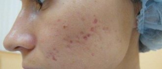
Of course, these hidden processes are reflected in the condition of the dermis: it becomes thin, dries out, and takes on a pale or grayish tint. The epithelium also loses its firmness and elasticity, as a result of which wrinkled networks appear on the skin, which gradually grow and acquire depth, turning into age-related folds. And the older a person is, the faster the pace of development of these negative processes. Age-related changes appear most quickly in the neck, face, décolleté and arms.
Thanks to cellular technologies, the aging process can be, if not stopped, then significantly reduced in intensity. Stimulation of fibroblast cells fights aging from the inside and that is why procedures using cells of this type are so effective.
Rules of rejuvenation: areas of responsibility for anti-aging
Dermal cells with the prefix “fibro-” are known in two types, and each of them performs its own function in maintaining youthful skin. Fibrocytes are more mature, stable cells; their task is to control the structural condition of the skin in certain areas. It is fibrocytes that synthesize glycosaminoglycans to create the extracellular matrix, and the most famous of them is hyaluronic acid. Fibrocytes also produce different types of collagen, and depending on the type and severity of skin damage (whether it is a sunburn, hard peeling or age-related changes), the ratio of different types of collagen produced changes.
Fibroblasts are younger cells that can move between layers of skin and transform into other types of cells if damaged skin needs it to regenerate. The skin owes fibroblasts processes that tighten the edges of the wound and restore the integrity of the damaged epidermis.
Despite the different characteristics and functions of cells with the prefix “fibro-”, they are all commonly called fibroblasts. But even in different layers of the skin they act differently, synthesizing the protein compounds it needs.
In the upper, papillary layer of the dermis, fibroblasts produce mainly collagen types 1 and 3, although there are also types 12 and 16, and the collagen fibers are thin and relatively randomly located. The proteins tenascin-C and decorin are also expressed here. In the deeper, reticular layer of the dermis, the production of type 3 collagen predominates, but there is also type 4. Collagen fibers are thicker and more ordered than in the papillary layer. In addition, elastin, tenascin-X, and versican proteins are synthesized in the reticular layer.
Due to differences in the work of fibroblasts, the papillary layer determines the appearance of the skin and its elasticity, and the reticular layer determines its strength and elasticity.
Fibroblast injections: the essence of the procedure
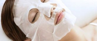
The essence of the procedure is quite simple: young connective tissue cells are transplanted into the dermis. Moreover, the method uses one’s own cells, which allows not only to avoid a rejection reaction, but also to maximally accelerate the processes of renewal and regeneration of the skin. The result of the procedure will be a noticeable improvement in complexion, reduction of wrinkles of all types (from facial to deep), deep hydration of the dermis, healing of scars and post-acne.
The main advantage of the technology is that fibroblast injections are highly effective and retain their beneficial qualities for a long time.
The results from the procedure will last for at least six months!
And all this time, collagen, elastin and hyaluron will be actively produced in the dermis and in the required quantity.
The cells for injection are obtained from the patient's dermis, a small piece of which is usually taken from the navel area or from the area behind the ears. It is in these areas of the human body that the skin is least susceptible to the negative effects of environmental factors. This piece is carefully examined and processed in the laboratory, stimulating the growth of young fibroblast cells. On average, cell cultivation takes a month to a month and a half, after which the grown cells are injected into the clinic patient’s area in need of correction.
A pronounced result can be expected after the first autotransplantation session, but to achieve a significant effect, cosmetologists advise taking a two-week course. Six months after the procedure, the depth of age-related changes in the area around the eyes is reduced to almost ninety percent, around the mouth - to fifty-five percent, on the neck and chest area - to ninety-five percent.
The role of fibroblasts in the process of epithelial proliferation
Proliferation of epithelial cells is impossible without fibroblasts. In these cells, increased protein production occurs during epithelial proliferation. This can be seen by increasing pyroninophilia in the cytoplasm. This indicates the accumulation of ribonucleoproteins, with the help of which a matrix for tropocollagen is formed. As a rule, epithelium with signs of proliferation includes mature forms of connective cells - fibrocytes, which are obtained from transformed fibroblasts. At the final stage, proliferation of epithelial cells is characterized by the formation of waste products of fibroblasts. Initially, these products are argyrophilic fibers, which subsequently participate in the formation of collagen fibers. Collagen structures provide a clear demarcation of the source of inflammation from normal tissue. Subsequently, collagen fibers grow and gradually replace the epithelium.
BENEFITS OF CELL THERAPY
The fibroblast rejuvenation technique has long enjoyed deserved popularity not only in Russia, but also in the UK, Switzerland and the USA. Extensive experience in using the technology allows us to assert that the truly fantastic rejuvenating effect of the procedure lasts for approximately 7 years. No other non-surgical technique can boast such a long-lasting effect.
The first result of the introduction of autologous fibroblasts cultured in the laboratory can be seen within a month after their first transplantation. Starting from this moment, over the course of 18-24 months, the number of cells responsible for the beauty and youth of the skin will steadily increase. They will work tirelessly to not give old age a single chance. The visual manifestation of this process is an increasingly youthful and blooming appearance.
Advantages of the technique:
- The firmness and elasticity of the skin increases. Soft and delicate to the touch, the skin is filled with life-giving moisture and radiance day after day.
- The surface of the face, neck, décolleté, and hands is smoothed. According to data obtained during clinical studies, just one month after the introduction of fibroblasts, wrinkles are reduced by approximately 14%, and after a year - by 46%.
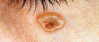
- Pigment spots, which visually add several years of age at once, fade and then disappear completely.
- The skin becomes dense and full. A year after the procedure, the thickness of the dermis can increase to 65% (no other method can provide this indicator).
- The patient no longer experiences a feeling of tightness and dryness of the skin.
- The face acquires a pleasant fresh color and is covered with a beautiful and healthy blush.
- The use of autologous fibroblasts implies a 100% guarantee of the absence of allergic reactions and rejection. After all, during the procedure the patient’s own biological material is introduced.
- Another important advantage of the technology is its excellent compatibility with other cosmetology techniques. Moreover, if you introduce cultured fibroblasts before laser resurfacing or other plastic surgery, the effect of the latter will be more pronounced and lasting.
- The advantage of the procedure is the absence of withdrawal effects. The fact is that, firstly, rejuvenation with fibroblasts is a natural, not an artificial process. Secondly, after transplantation of fibroblasts, the decrease in their number in the body does not occur sharply, but very slowly.
- The rejuvenating effect of fibroblasts lasts for years and years. Traces of the past years literally melt away before our eyes. Imagine how much money you can save during this time on multiple-use cosmetic procedures.
PHOTOS BEFORE AND AFTER FIBROBLAST REJUVENATION
In the Photo Gallery section you can see photos before and after cell therapy .
AFTER THE PROCEDURE OF AUTOLOGICAL FIBROBLASTS INTRODUCTION
Mild redness and slight swelling of the treated area are normal and may occur for several hours after cell therapy and then disappear without a trace.
Transplanted fibroblasts begin to actively produce components of the intercellular matrix, thereby enhancing microcirculation in the skin, regenerating the collagen framework, and increasing the thickness of the dermis. After 6-8 hours, the natural process of self-rejuvenation of the dermis is in full swing: turgor is restored, skin elasticity returns, and nutrition of cells and tissues is enhanced. After some time, small wrinkles disappear as if by magic, and deep ones become noticeable much less. Some patients see the rejuvenating effect of the procedure after 2 weeks, but more often the effect becomes noticeable after 1 - 1.5 months. Gradually, the result becomes more pronounced and reaches a maximum after about a year. A reflection is looking at you from the mirror, which you probably missed, since you last saw it 10-15 years ago.
Patients of the Abrielle Clinic have the tempting opportunity to enter into an agreement with the Stem Cell Bank and preserve their own fibroblasts in liquid nitrogen for an arbitrarily long period of time. Individually stored cultured “youth cells” can be used as needed to restore a youthful glow after 5, 10 or 15 years. In other words, cryogenic storage of fibroblasts is a unique chance to preserve unfading beauty for many years, taking care of it today.
Do you want to collect a rich harvest of compliments every day? Do you want to regularly hear: “What gorgeous skin you have! Tell us the secret, what is the name of your cosmetics? Take advantage of the revolutionary discovery of modern cosmetology and academic science - discover a technique that allows you to restore youth by influencing the body at the cellular level. Fill your own skin with strength and energy, eliminate the very cause of aging. Give your skin the opportunity to renew itself from the inside.
SPRS therapy: 12 steps to youth
- Arrival at the clinic.
- Receiving advice and undergoing examinations.
- Elimination of all possible contraindications.
- Collection of biomaterial from the behind-the-ear area.
- Transportation of material to the biological laboratory.
- SPRS diagnostics.
- Drawing up a “skin passport” and developing a treatment program.
- Creation of the drug.
- Transportation of the drug to the clinic.
- Storage of fibroblast culture.
- Administration of the drug.
- The final stage of therapy.
Skin rejuvenation with your own fibroblasts.
The essence of SPRS therapy® is that a piece of skin with a diameter of 4 mm is taken from the patient’s ear (where the skin is least damaged by UV rays) and a fibroblast culture is obtained from it. In the skin of every person, regardless of age, fibroblast precursor cells are present. In culture, they are able to form colonies - clones consisting of tens and even thousands of fibroblasts, developed from a single precursor cell. This means that in a person of any age, from a small fragment of skin, you can obtain the number of fibroblasts necessary for therapy. We are not exaggerating when we talk about “any age”: in the skin of even a 95-year-old person there are up to 14% active fibroblasts!
As it turned out, by culturing cells it is possible to obtain a very interesting individual characteristic of the skin, on the basis of which one can calculate the value of the regenerative potential of fibroblasts (that is, their ability to maintain tissue self-regulation and restore it when damaged), on the basis of which a conclusion can then be made about the regenerative potential of the skin in general, and, therefore, its ability to self-heal. The magnitude of this potential is individual for each person, it does not depend on his age. Taking this indicator into account, it is possible for each patient to develop his own individual program for skin renewal and restoration (the so-called “Skin Passport®” and “SPRS program”).
What is it for? Everyone is familiar with this situation: two patients of the same age undergo the same procedures, then one is completely delighted, and the other complains that there is no point. “Skin Passport” helps predict the result, find out who will easily get an excellent effect, and who needs to undergo preparatory procedures. This is the essence of the individual SPRS program.