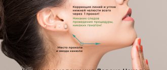
The nasolacrimal trough is a depression that forms at the border of the cheeks and the area under the eyes. Many factors can provoke its appearance, but the most common of them are age-related changes and genetic predisposition.
Many women over 40 have this deficiency, but it can often be seen on very young girls. Next, you will find out when correction of the nasolacrimal trough
and how it can be done, as well as other reasons for its appearance.
Why does a nasolacrimal trough form?
A cosmetic defect can occur in middle-aged and elderly women, but it also occurs in girls under 30 years old, which is due to the characteristics of the skin and other factors.
The furrow is a slight depression formed in the area of the inner corner of the eye, and continues in the direction of the arc that forms the cheekbones. It is also called a deep wrinkle or fold.
There are several reasons that can provoke the problem:
- A sharp change in body weight in one direction or another. In this case, the skin sags or stretches, which certainly leads to a loss of elasticity.
- Bad habits. Women who abuse alcoholic beverages and tobacco products are more likely to notice the appearance of folds.
- Excessive physical activity and fatigue.
- Professional activities associated with contact with toxic substances. Typically, such work has a negative impact on the skin.
- Age-related changes in the skin associated with a decrease in the production of collagen and other components that maintain the elasticity of the skin.
- Skull injuries.
- Disorders of the heart and kidneys, causing stagnation of blood and fluid.
- Severe brain pathologies.
- Regular insomnia associated with psychoemotional disorders.
- Diseases of the endocrine system, for example, thyrotoxicosis, hypothyroidism or diabetes.
One of the common reasons is considered to be improper care of the skin of the face. An unsuitable cream, scrub or lotion can cause tissue swelling and other problems that ultimately lead to the appearance of creases.
The nasolacrimal and palpebromalar grooves are one of the most difficult areas to correct. The complexity of their anatomical structure and the fear of making a mistake stop cosmetologists from injecting. Many of us forever refuse to work in these important areas of the face from the point of view of rejuvenation. Therefore, I would like to once again discuss important nuances that can be useful for both beginners and experienced doctors.
Anatomy of the zone
Since no invasive manipulations are most often performed in the projection of the palpebral part of the orbicularis oculi muscle, let us recall the layer-by-layer anatomy in the projection of its orbital part.
The skin of this area is thin, the underlying superficial fat is absent both on the palpebral part and below the bony edge of the orbital foramen for approximately a centimeter. This structure does not allow adequate filling of wrinkle lines here even with the most flexible fillers in their rheological properties.
Before
Subcutaneous fat is represented by superficial infraorbital compartments. Their lower border is the zygomatic ligament, the upper is the ligament that supports the orbicularis oculi muscle, the surface is represented by the skin, and the bottom is the orbicularis oculi muscle.
The connective tissue of the zygomatic ligament, with which it is attached to the skin, is a frequent, vertically oriented trabeculae located exactly along the route of lymph drainage from the lower eyelid to the submandibular lymph nodes, which creates an obstacle to its outflow. Due to metabolic disorders, partial reduction of the tone of the orbicularis oculi muscle by botulinum toxin and other factors, chronic lymphatic stagnation in the form of a malar hernial sac is formed in this area.
The next layer of tissue here is the orbital part of the orbicularis oculi muscle, the free edge of which reaches the zygomatic ligament. Immediately at the medial canthus of the eye, it is connected to the periosteum of the zygomatic bone. Its periosteal attachment continues for 5 mm. Next, the muscle is fixed to the bony edge of the orbit with the help of connective tissue located perpendicular to it - the ligament that supports the orbicularis oculi muscle.
Below this is the deep infraorbital fat compartment, known by the English acronym SOOF. It is present only on the zygomatic bone and is tightly attached to it. Since the zygomatic bone is more convex in the anterior plane of the face, in comparison with the upper jaw bone, nature rationally placed the SOOF only on its periosteum so that the inner surface of the orbicularis oculi muscle slides along it during contractions. Immediately below the orbital foramen on the upper jaw there is no deep fat. The lower bony edge of the orbit is represented by the maxillary and zygomatic bones anastomosing with each other.
The periosteum in the anastomosis zone between them grows as a protrusion into the anterior plane of the face and covers the highest point of the “canine fossa”, in which the infraorbital foramen is located with the neurovascular bundle emerging from it. In addition to the periosteal protrusion above the recess of the “dog fossa,” there is another factor that ensures the safety of the infraorbital neurovascular bundle - its channel is located not perpendicular, but at an angle relative to the surface of the periosteum.
After
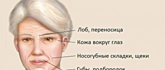
Anatomical and aesthetic nasolacrimal groove
The muscle fibers in this area are thicker, well filled with blood and have a burgundy color. Both deep and superficial fats are absent in this area, the surface of the skin is deepened and the burgundy color of the muscle shines through the thinness of the tissue.
In the literature, the nasolacrimal groove is described as a zone of deepening and absorption of light, starting from the medial canthus and continuing to the midpupillary perpendicular.
The so-called aesthetic nasolacrimal groove from an anatomical point of view is a canal, the upper boundary of which is the periosteal attachment of the orbicularis oculi muscle, the lower boundary is the septa of the angular vein, from above it is covered by the orbicularis oculi muscle, and its bottom is represented by the periosteum of the upper jaw. The visual aspect of the palpebromalar groove is ensured by the fact that the external malar fat does not reach the bony edge of the orbit and ends a centimeter earlier. Above the palpebromalar groove, the skin lies directly on the orbicularis oculi muscle, so its surface is deepened. The visual appearance of the palpebromalar groove adds contrast between the skin of the anterior zygomatic zone, where it is thick and has ducts of the sebaceous glands, and the skin of the lower eyelid, which is thin and without ducts of the sebaceous glands.
The morphological nasolacrimal groove exists from birth. It is a periosteal attachment to the orbicularis oculi muscle, approximately four millimeters in length.
Detailed description of correction tasks
It is impossible to overestimate the importance of correction of infraorbital grooves in the overall concept of facial rejuvenation.
The eyes are the central zone of visual attraction, and any morphological and involutional changes in it distract the interlocutor from the central focus of the gaze.
It is worth taking into account that the aesthetic objectives of correction are much broader than the anatomical ones. From an anatomical point of view, you should simply fill the recesses of the lower orbital grooves. From an aesthetic point of view, it must be remembered that the infraorbital region from below passes into the anterior zygomatic region, and from the side into the temporal region, and the transition between them should be smooth.
So, let's set the tasks. It is necessary to reduce the recesses of the nasolacrimal and palpebromalar grooves, visually shorten the lower eyelid, disguise the palpebral and malar hernial sacs and restore a smooth transition from the eyelid to the cheekbone in the anterior and lateral angles.
In normal practice, the doctor is faced with the need to simultaneously correct the lower orbital grooves and the zygomatic region. A continuation of the nasolacrimal groove is the nasobuccal groove. If you inject only the nasolacrimal groove, then against its corrected background the nasobuccal groove looks even deeper.
It should be remembered that the normal configuration of the infraorbital grooves from an aesthetic point of view presupposes the presence of a small nasolacrimal groove and the absence of a palpebromalar groove. If only the nasolacrimal is corrected, then the smoothness of the eyelid junction is restored anteriorly, but is not restored
from a side perspective. The configuration of the infraorbital grooves is disrupted, the nasolacrimal groove is absent, and the palpebromalar groove is visually emphasized and highlighted.
When injecting the filler, lidocaine automatically enters the tissues and creates infiltration anesthesia in them. Subsequent injection of the drug into the nasolacrimal groove against this background feels much less painful.
Correction methods
Conventional practice requires simultaneous correction of the anterior zygomatic and infraorbital areas. The task of correction in the anterior zygomatic zone is to restore its uniform smooth convexity. In this case, the gel is injected first into the deep medial buccal compartment, and then into the nasolacrimal and palpebromalar grooves.
In the presence of lidocaine-containing drugs, this sequence seems to be the most rational.
The instrument of choice for injections into the nasolacrimal groove is a cannula that is not too long (4 cm) and fairly thick (25G) in diameter. The nasolacrimal groove is one of the most dangerous areas for correction. Normally, the angular vein and the superior branch of the infraorbital artery always pass along this route.
Anatomical variations in thirty percent of cases ensure the passage of the angular artery itself in this zone and in a small percentage of cases its duplex.
It is known that the angular artery anastomoses here with the internal carotid system (with the supoatrochlear artery) through the dorsal nasal artery. In retrograde embolism, through this artery the embolus enters the internal carotid system and creates an occlusion in the central retinal artery, which causes blindness.
The entrance hole should be selected on a perpendicular line through the lateral canthus above the septum of the angular vein, but below the zygomatic ligament. The trocar should only pierce the skin and, to avoid bleeding, advance into the tissues with a cannula. The cannula should be located under the orbicularis oculi muscle. The latter here represents the SMAS level, so the insertion tool should not lift the overlying tissue when attempting to do so. The level of the periosteum in the vestibule of the orbit changes, first rising and then descending. When passing into the nasolacrimal groove, the cannula must follow the path of the periosteum, otherwise you can pierce the orbicularis oculi muscle with it and introduce the gel superficially.
The test for the correct level of insertion of the cannula should look like this: when trying to move it up, it should not move, resting against the periosteal attachment of the orbicularis oculi muscle; when trying to move it down, it should rest against the septa of the angular vein, which is the upper border of the nasolabial compartment. If you puncture it, the injected gel creates a protrusion in the form of a roller along the septum of the angular vein, and the filling of the groove does not occur. When trying to lift the cannula up and protrude the overlying tissues, the overlying orbicularis muscle prevents this from being done. And finally, when trying to press on the bottom of the canal of the nasolacrimal groove, the cannula rests on the periosteum of the upper jaw.
To ensure that lidocaine works, the gel should be injected first antegrade, slowly moving towards the medial canthus, and then retrograde until complete correction. It often happens that the cannula goes in correctly, but too deep, and there is no filling of the sulcus during insertion. In this case, you need to go back a little and try to pass the cannula just under the bottom of the orbicularis muscle, still under it, but not supraperiosteally.
If you move half a centimeter down along the route of the nasolacrimal groove, the supraperiosteal attachment of the orbicularis oculi muscle ends, and the cannula, if necessary, can easily pass to its suspensory ligament. Often, immediately under the suspensory ligament, the SOOF is not yet strong; here its volume can be replenished by introducing filler and making the transition from the eyelid to the cheekbone smoother.
If in the nasolacrimal groove, due to anatomical danger, the instrument of choice is a rigid cannula, in the palpebromalar groove the injection can be carried out with a needle. If you turn the cannula upward from the nasolacrimal groove towards the suspensory ligament, it becomes difficult to bend over the convexity of the orbital vestibule. Closer to the lateral corner of the eye, the ligament of the orbicularis oculi muscle begins to attach below the bony edge of the orbit and, on the lateral edge of the orbit, fuses with the upper one, forming the lateral orbital thickening.
When correcting the palpebromalar groove, the needle should be inserted at an acute angle until it touches the periosteum, being positioned just below the bony edge of the orbital foramen, but above the ligament that supports the orbicularis oculi muscle. Otherwise, you can pierce the palpebral hernial sac that often protrudes from the orbit at the top or insert it into the underlying malar sac. Correct insertion in this area allows you to hide the palpebral and malar hernial sacs and restore a smooth eyelid junction in its lateral part.
Conclusion
If you base your practice on detailed knowledge of the anatomy of the area and use properly selected instruments and materials, the risk of undesirable consequences of correction can be minimized.
On the contrary, the aesthetic results for the patient should be made as satisfactory as possible.
Author: Margarita Egorova
Magazine: Appearance. Esthetic guide №2(25)
The effectiveness of correction of the nasolacrimal groove with fillers and hyaluronic acid
Filling the nasolacrimal trough with fillers is considered the most accessible and simple procedure for those who are contraindicated for surgery or have other obstacles.
Fillers are made based on hyaluronic acid and other components. They are a gel-like mass that fills the voids under the skin and leads to the alignment of its relief. In this case, there is no need for an operation, the procedure takes a minimum of time, and its results are long-term.
It not only smoothes out the skin, but also stimulates metabolic processes and blood circulation, which improves color and eliminates redness. Many women feel discomfort when furrows form because the skin under the eyes becomes sensitive, thins and dries out quickly.
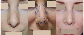
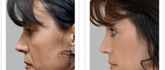
Filling the nasolacrimal groove with fillers is the most effective and safest option for correcting this area of the face. Photos before and after the procedure.
After the procedure, all these problems disappear, which also proves its effectiveness and safety if carried out correctly.
Correction
The procedure will be useful for those who:
- Has bad habits (tobacco and alcohol use).
- Leads a passive lifestyle.
- Wears glasses for a long time.
- Has chronic fatigue.
- He suffers from insomnia, as a result of which it is imprinted on his face.
- Has certain diseases and pathologies.
Contraindications for nasal tear correction:
- Diabetes.
- If this procedure has already been performed.
- Viral and infectious diseases.
- Oncological diseases.
- Breastfeeding and pregnancy.
- Chronic disease processes.
- Drug intolerance.
- Taking antibiotics.
- Hemophilia.
Preparation
Preparation for correction of the nasolacrimal trough is simple. If you have no contraindications to this procedure, then you simply need to refrain from tanning for a week. If your skin is prone to hypersensitivity and your blood vessels are fragile, then to avoid bruising and hematomas, take a course of ascorutin.
In any case, before the procedure you will consult with a cosmetologist and he will give you recommendations before the procedure.
Nasolacrimal insertion procedure
The procedure itself is quite quick and easy to perform. Filling a nasal tear with filler takes at most 10 minutes. And if you add to this time all the stages of preparation, including the effects of anesthetics, then expect to stay with the cosmetologist for about an hour and a half.
The procedure itself goes as follows:
- At the preliminary consultation, the cosmetologist will tell you in detail how exactly you need to prepare for the procedure.
- Next, he will select the filler that is right for you.
- The facial skin is thoroughly cleansed of excess makeup.
- Next, the cosmetologist marks with a marker the places where filler needs to be injected. This is necessary so that the swelling does not confuse you.
- An anesthetic cream is applied. For this cream to start working, you need to lie down for about 30-60 minutes.
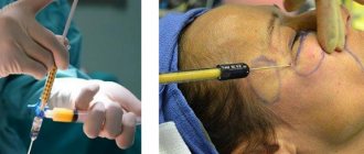
- Next, the specialist injects the filler into the previously marked areas. The filler is injected in small portions using zigzag movements.
- At the end, the cosmetologist distributes the gel under the skin with his hands, giving the desired shape and smoothing out the transition areas of the gel.
We invite you to watch a video about how the procedure for correcting the nasolacrimal groove with filler is carried out:
After correction
After the procedure, you must adhere to several rules so that the result pleases you for a long time and healing proceeds without complications:
- The very first rule is to refrain from visiting the bathhouse, sauna and solarium for two weeks.
- Also, during the first weeks, try to avoid direct exposure to sunlight. Wearing sunglasses is also contraindicated.
- For the first time, you will have to give up any physical activity and visiting the fitness room.
- Do not consume alcohol or tobacco.
- Do not touch or rub the places where the procedure took place.
Can it be installed at home?
Under no circumstances should you attempt to correct a nasolacrimal trench yourself at home unless you have a medical background. If you inadvertently damage blood vessels in the tissues, this can lead to consequences such as bruising under the eyes, bruises and dead skin. The facial nerve can also be easily hit, resulting in paralysis. Therefore, this procedure should be done by specialists in this field in specialized salons .
Complications and swelling
Before agreeing to correct the tear trough, it is necessary to thoroughly study all the intricacies of this procedure. Compare all the risks and possibilities of complications with the state of your health, and only then agree to this manipulation.
- It happens that the filler substance can compress small capillaries, resulting in swelling. During the procedure, the vessels can be overlooked, which is why this problem occurs. You don’t need to do anything for this; the swelling should go away on its own.
- Before the procedure, find out what concentration of acid the doctor recommended for you. If you get the wrong type of filler, it may spread and go somewhere else.
- It happens that the filler is injected in excess quantities, in which case, with any facial expressions, traces of it on the face will be noticeable, which greatly spoils the aesthetic appearance. in this case, you can correct it by injecting Longidase, which will help the gel dissolve.
- The filler is injected only under the skin. If it is inserted deeper, a scar may appear in this place.
- If the procedure was not carried out by a professional specialist, then there is a possibility that the filler substance will appear in nodules or lumps.
Most of the unwanted complications are associated with improper work of the master . The procedure for introducing filler into the nasolacrimal groove, about which patients leave positive reviews, is quite complicated, so you need to choose a professional technician who has extensive experience. It is better to give preference to a more expensive clinic, but with a good reputation. You should not forget to read reviews about the establishment in order to understand what awaits in the future.
Contraindications for correction
Despite the relative safety and effectiveness of the manipulation, it is not always permitted.
Some contraindications must be taken into account:
- Intolerance to drugs used in furrow correction.
- Any malignant neoplasms in the body.
- Eczema and psoriasis in the acute stage.
- Chronic dermatitis, urticaria with regular relapses.
- Damage to the skin in the furrow area.
- Purulent lesions on the skin in the eye or nasal passages.
In each case, the specialist individually makes a decision on the advisability of the procedure.
Contraindications
Injection of filler into the nasolacrimal groove is prohibited if:
- tendency to swelling of the face;
- hernias under the eyes;
- bearing a child;
- breastfeeding;
- inflammation on the face in the place where injections are planned;
- chronic pathologies;
- diseases in the acute phase;
- oncology;
- epilepsy;
- autoimmune diseases;
- tendency to form keloid scars;
- hemophilia.
Fillers injected into the nasolacrimal groove effectively cope with problems such as blueness and puffiness under the eyes, wrinkles and sagging skin. Each cosmetology clinic offers its own range of drugs, each of which is selected individually, taking into account the existing problems and characteristics of each patient’s body.
Fillers can also be injected into other areas. You can read how they are used for lips, cheekbones, private parts, nose, chin, face, forehead, nasolabial folds, cheeks.
Comparison of the technique with other correction methods
Correction with fillers has its advantages and disadvantages. A woman should familiarize herself with all the manipulations that can eliminate the furrow in order to understand which one is right for her.
| Name of procedures | Peculiarities | Effect duration |
| Surgical intervention | There are 2 main procedures that require surgery - lipolifting and cutting off adipose tissue. The first procedure involves taking adipose tissue from different parts of the patient’s body and then filling the nasolacrimal fold with it. It is performed under local anesthesia. The second involves cutting off fatty tissue in the area of the furrow, if it is quite deep and combined with bags under the eyes. The manipulation is performed under general anesthesia. | The effect of lipolifting lasts for 3 years; after excision, the specialist gives a guarantee for 10 years. |
| Hardware techniques | Today, lifting with ultrasound and radio waves is considered the most popular. Both manipulations are carried out in courses, the number of procedures depends on the severity of the furrow. The patient does not require pain relief. Thanks to the healing effect, metabolism in the fold area is stimulated, and its own collagen is formed, which fills the space. As a result, the fold is smoothed out. | The effect of such manipulations lasts no more than 3-4 months. |
| Injection method of correction | Fillers based on hyaluronic acid and other components fill the voids in the area of the nasolacrimal groove, which leads to its smoothing. | After manipulation, the result lasts for 6-8 months. |
The disadvantage of correction with fillers is the short-lived effect. However, taking into account the risks that the patient is exposed to during surgery, this manipulation is considered more gentle.
Technique of drug administration
Before a date is set for injection of fillers into the nasolacrimal groove area, the patient must visit a cosmetologist. This initial consultation should identify existing contraindications and the hidden course of infectious and inflammatory processes. The doctor will assess the upcoming amount of work, decide on the choice of filler and the amount that will need to be injected. Then the cosmetologist acts according to the following algorithm:
- Cleanses the skin of dirt, dust, decorative cosmetics. Treats the area of the nasolacrimal groove with an antiseptic solution.
- Apply cream or gel with anesthetic to the face. This step is performed only if the filler does not contain an anesthetic. The gel should be removed 20 minutes after application.
- Injections are performed at a certain distance from each other. Each puncture is wiped with a sterile cloth to prevent the infectious agent from penetrating into the deep layers of the dermis.
After the required amount of the drug is administered, the doctor performs several soft, gentle massage movements. This promotes a more even distribution of the filler.
The last stage is applying a soothing, cooling mask to the face.
Watch this video about what the procedure for correcting the nasolacrimal trough is:
Types of drugs
Filling the nasolacrimal groove today is carried out using drugs from 3 groups. Each group has its own advantages and disadvantages.
Synthetic
Preparations from this group were created in the last century; they mainly contain paraffin and silicone. The main advantage of the funds is considered to be low cost. They provide a fairly long-lasting result and do not react with the patient’s skin cells.
However, synthetic fillers have a significant drawback. Once introduced into the voids, they are able to migrate to other areas, which can lead to the appearance of atypical defects on the face. An additional disadvantage of such drugs is the increased risk of developing an allergic reaction after administration.
Biosynthetic
Such products are considered 2nd generation drugs because they are based on synthetic ones, but are a more preferable option due to better compatibility with skin cells. Another advantage is the ability to persist in the human body for about 2-5 years.
Such products contain synthetic polylactic acid, as well as polycaprolactone and calcium hydroxyapatite. An important disadvantage of biosynthetic drugs is blockage of blood vessels and swelling of tissues. In this case, signs may appear immediately after administration or after some time.
Biodegradable
Popular fillers are made on the basis of a special substance, which, after injection, stimulates the natural processes of smoothing the skin. At the same time, the likelihood of adverse reactions is minimal, and the effect lasts from 8 to 10 months, which is almost the maximum period when using fillers.
Most often, the main substance is hyaluronic acid, which is normally produced in the human body and has unique properties. Another advantage of such drugs is the ability to be eliminated from the body naturally. Today, biodegradable fillers are considered the best and are used most often, as they guarantee results.
Preparation for the procedure
Preparing for the procedure does not take much time.
After selecting the appropriate drug, the following steps are performed:
- The specialist places the patient on the couch and puts a disposable cap on her head.
- Using special products, the skin is cleansed from impurities and excess fat.
- After this, the cosmetologist makes markings along the line of the furrow.
- An anesthetic is injected into the required areas, but more often a special cream with anesthetic components is used.
Before performing all the manipulations, the specialist tells the patient about the procedure, its duration and effectiveness. She should have an idea of all the actions of a cosmetologist.
How to make a correction
Filling is carried out after the anesthetic begins to act. The specialist begins the injection of filler or pure hyaluronic acid along the line of the nasolacrimal groove, marked at the preparatory stage. When using hyaluronic acid, the algorithm of actions is no different.
Using a disposable syringe with a miniature needle, a specialist injects a drop of gel at an equal distance, which allows the product to be evenly distributed. The punctures are shallow, usually the patient does not feel discomfort.
After treating the entire crease line, a gentle massage of the treatment area is carried out in order to distribute the gel and prevent its accumulation in one area under the skin. The entire manipulation lasts no more than 1 hour.
Many clinics offer women a soothing mask, which is applied after the filler is injected to cool the skin and prevent the development of inflammation.
Recovery time, features of the course
The duration of recovery after manipulation varies from woman to woman. Don't expect an immediate effect. It usually takes 10 to 14 days for the skin to completely recover and swelling and redness to disappear.
Throughout this entire period, it is recommended to follow a diet, namely, exclude salty, spicy and sour foods from the menu. It is important not to drink alcohol or take strong drugs, such as painkillers.
In addition, it is strictly forbidden to take a hot bath or sunbathe near a pond or in a solarium . You should not visit a bathhouse or sauna to avoid the possibility of increased swelling. If you follow the recommendations, the recovery period proceeds favorably.
Care after correction
If proper care is taken after correction, the likelihood of a furrow forming in the same place increases, so you should follow the cosmetologist’s recommendations.
Recommendations:
- The daily cream should be suitable for the patient’s skin type and age.
- When using decorative cosmetics, it is important to cleanse the skin with micellar water before going to bed.
- Creams with a cooling effect can be used for 2 weeks after correction.
- It is useful to use a cream for the area around the eyes containing hyaluronic acid. He will maintain the result.
You should not rub the area where the furrow was or massage or pull the skin so as not to provoke complications.
Possible complications and methods of their treatment
Filling the nasolacrimal trough with fillers often provokes complications. They can be minor or dangerous, so you should not ignore the symptoms.
Edema
Normally, tissue swelling disappears 3 days after the procedure. If this does not happen, you should visit a doctor who will determine the cause of the disorder and prescribe treatment. Usually a specialist prescribes an ointment that helps eliminate swelling. The most commonly used is Heparin ointment, which is applied 2 times a day in a thin layer for 3-6 days.
It is worth noting that the instructions do not suggest such use, but the experience of many patients proves its effectiveness. In most cases, additional therapy is not required.
Pain
Pain in the area where the gel was inserted may persist for several weeks after the procedure . The symptom is caused by skin damage or uneven distribution of the drug. Often pain is a reaction of the skin to the diameter of the needle.
If the pain is present only when touched, there is no need to worry, it will disappear after a while. If discomfort is constant, you should consult a doctor.
He will prescribe non-steroidal anti-inflammatory drugs, such as Indomethacin or Ibuprofen.
The drugs help eliminate pain, swelling and reduce the temperature in the affected area. This will ease the woman's condition. You need to take 1 tablet 2 times a day for no more than 5 days in a row.
Inflammation
The inflammatory process after correction in most cases is associated with the body’s reaction to a foreign substance. If swelling and redness does not disappear after 3 days, the symptoms worsen and other manifestations occur, you should consult a doctor.
Treatment usually involves taking antibiotics, such as Azithromycin.
The product helps to destroy microorganisms in the area of inflammation and is highly effective even when taking small doses.
The patient needs to take 1 tablet in the morning and evening for 3-5 days. If necessary, antipyretics are prescribed, for example, Paracetamol, as well as painkillers (Ketorol).
Necrosis
Necrosis is considered the most dangerous complication. It is the necrosis of tissue areas as a result of disruption of innervation and blood supply. In this case, swelling develops, redness and bruising appear.
After some time, the areas under the eyes turn black. This condition is treated by surgical excision of areas of dead tissue followed by antibiotic therapy. After surgery, the likelihood of facial expression disturbances increases.
Consequences and complications
Main consequences and complications:
- Poor tissue survival;
- Clumping of adipose tissue under the skin;
- Formation of hematomas and edema;
- Infection of the injection area;
- Formation of compactions;
- Tissue fibrosis in the injection area;
- Uneven distribution of tissue under the skin.
Side effects in the form of hematomas and swelling go away on their own quite quickly - usually after a week there are no traces left.
The best drugs for filling the nasolacrimal trough
Filling the nasolacrimal groove is carried out using drugs with different compositions and properties.
There are tools that are used most often:
- Juvederm – the most popular products containing hyaluronic acid. This fills the furrow and stimulates the skin's natural smoothing processes. The drugs are effective, rarely provoke inflammation and necrosis, and many contain lidocaine to reduce pain. The main disadvantage of the funds is the increased risk of developing allergies.
- Teosyal redensity 2 are popular fillers, the composition of which includes hyaluronic acid supplemented with glycerol. The products are safe, retain their effect for at least 10 months and are well tolerated by patients. The only drawback is the high cost.
- Belotero soft – a fairly effective filler, which contains hyaluronic acid salts, but a minimal content of sodium hyaluronate. The product provides a long-lasting effect and rarely leads to complications. It also stimulates natural tissue repair processes and improves skin color. The drug has no disadvantages.
- Stylage S is a new generation French filler that provides long-lasting results and does not provoke allergic reactions. The composition contains high-quality hyaluronic acid, as well as other components that ensure effectiveness. In addition to lifting properties, the product has an antioxidant effect. The only drawback of the product is its high cost. However, the price is justified considering that the gel is packaged in disposable, convenient syringes, packaged in moisture-proof blisters.
There are many more drugs for correcting the nasolacrimal trench. Each of them has its own advantages and disadvantages. The choice is made by a specialist, taking into account the individual characteristics of each patient.
How to prevent tear trough formation?
Everyone knows that any problem is always easier to prevent than to treat. Therefore, prevention of tear trough formation should be started immediately. And if there is a hereditary predisposition, then this can be revealed by looking at your relatives - then you will have to work even more actively. Even if a problem has arisen, a list of innovations in your usual way of life will help correct the situation:
- proper nutrition - you need to eat more vegetables and fruits, less sugar, salt and smoked foods;
- regular physical activity (you don’t need to buy a gym membership at all, just a 30-minute leisurely walk is enough: this will have a positive effect on metabolic processes);
- correct drinking schedule (750 ml should be drunk before noon, the rest of the time the figure should be increased to 2 liters, but stop drinking an hour before bedtime);
- moisturizing your facial skin with creams that suit your age group;
- gymnastics for the face;
- treatment of chronic diseases.
We recommend: How to perform non-surgical blepharoplasty with mesoeye c71?
Cost of the procedure in Russian clinics
The price of correction of the nasolacrimal groove varies in different regions of Russia. In clinics in St. Petersburg and Moscow, the price ranges from 11,000-14,000 rubles. depending on the drug used.
In regions, for example, Voronezh, Kursk or Saratov, the price of such a correction can be 9,000-10,000 rubles. When using synthetic fillers, the cost of manipulation is slightly lower.
Filling voids in problem areas is considered a fairly popular procedure today. The nasolacrimal groove is most often corrected, since it is in this area that in most cases a cosmetic defect appears.
To ensure that the procedure does not provoke complications, you should listen to the recommendations of the cosmetologist during the recovery period.
Reviews
The procedure has received fairly mixed reviews. Many received positive results that lasted for years. But no less people noted a large number of complications that spoiled the face from an aesthetic point of view. There were also cases of low tissue survival. Considering that the procedure is not cheap, this effect often caused angry responses from clinic clients, although the explanation for this factor is usually the individual reaction of the body.
This video talks about lipofilling of the nose of a famous actress: