- Why does a plaque form on the eyelid?
- How does she look
- How is it different from xanthoma?
- Who are the people at risk?
- Can it become malignant?
- How to treat: all methods
- Surgical removal
- Electrocoagulation
- Radio wave destruction
- Possible complications after removal
- How to prevent the formation of xanthelasma
- How to reduce the risk of relapse of the disease
Whitish-yellow plaques on the eyelids and around the eyes, usually flat or slightly raised above the surface of the skin, are xanthelasmd . Mostly middle-aged and mature women face this cosmetic defect. The defect can appear either singly or in groups of plaques. The most common location for xanthelasma is the inner corner of the eye on the upper eyelid.
The prefix "xantho-" means yellow.
Xanthelasma: causes
Xanthelasma
From a medical point of view, xanthelasma of the eyelids is a benign formation of a flat (less often convex) shape, having a smooth or wrinkled surface and a characteristic whitish-yellow and sometimes light orange tint. The diameter can be from 2 mm to 5 cm. As a rule, xanthelasma does not bother a person in any way, is painless, and is only a cosmetic drawback. If the size of the plaque is small, it does not interfere with the proper functioning of the eyelid.
The exact reasons why this neoplasm appears have not been established. However, in the course of a considerable number of clinical studies, a clear relationship between the defect and such factors of the condition of the adult’s body was revealed, such as:
- excess body weight;
- diabetes mellitus (lack of the hormone insulin, which causes a persistent increase in glucose levels in the blood);
- myxedema (a disease considered an extreme form of hypothyroidism in adults - a long-term and persistent lack of thyroid hormones);
- pancreatitis (inflammation of the pancreas), liver damage;
- lipoid nephrosis (dystrophic kidney damage);
- high concentration of cholesterol in the blood.
As for children, xanthelasma in them is a hereditary pathology such as xanthomatosis. Subsequently, disturbances occur in the functioning of the liver and the functioning of the cardiovascular system. Often this is also accompanied by the formation of cysts on the hands.
Xanthomatosis is the formation of numerous xanthomas on the surface of the skin.
Drug treatment
Eliminating xanthelasma with a laser only temporarily solves the problem, since it is not able to affect the root cause of the disease. The occurrence of this pathology is associated with hereditary predisposition, dysfunction of the liver and pancreas, obesity, and diabetes mellitus.
However, most often xanthomas on the skin of the eyelids occur due to lipid metabolism disorders. That is why its normalization is the main condition for successful treatment. For this purpose, the patient is prescribed:
- Statins are drugs that help reduce the concentration of cholesterol in the blood by inhibiting its synthesis (Atorvastatin, Rosuvastatin, Simvastatin).
- Bile acid sequestrants are medications that bind dietary fat, thereby reducing its absorption in the intestine (Cholesteramine, Cholestipol).
- Fibrates - reduce the content of low-density lipoproteins responsible for the formation of plaques (Fenofibrate, Ciprofibrate).
- Probucol - can reduce the level of various fractions of cholesterol, does not affect triglycerides. This drug can be combined with other lipid-lowering drugs.
The necessary medications, dosage and duration of treatment are selected by the therapist depending on the general condition of the patient, the results of his general clinical tests and lipid profile.
Xanthomas and xanthelasmas - what is the difference?
Histologically these are similar formations. The main difference between the two is that xanthelasma is rarely associated with elevated triglyceride (lipid) levels. Only in some cases does this defect manifest itself due to a violation of fat metabolism in the body. But xanthomas, in turn, are formed due to lipid metabolism disorders.
With xanthelasma, as a rule, the level of lipids in the blood is normal, and subsequently there is a slight increase in cholesterol concentration. Despite this, it is often called flat xanthoma.
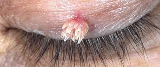
The difference can also be identified in relation to the places where neoplasms appear. If xanthelasmas are mainly plaques around the eyelids, having mainly a cosmetic defect, then xanthomas come in different types. For example, its appearance, such as tuberose, appears most often on the skin in the area of the knees and elbows. “Representatives” of another type – tendinous – “settle” on the extensor tendons of the feet and hands, as well as on the Achilles tendon. There are also: eruptive xanthomas are cluster-shaped formations of plaques that can appear on any part of the body; flat xanthomas.
Causes and symptoms of xanthelasma of the eyelids
The results of medical research suggest that the appearance of this neoplasm is caused by a malfunction in lipid metabolism. A comprehensive diagnosis of the owners of such plaques most often reveals pancreatitis, diabetes mellitus, liver disease and other diseases associated with increased cholesterol levels in the blood. This condition primarily affects women aged 50 years and older, although men can also develop it if they have certain health problems.
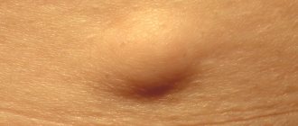
The neoplasm appears on the skin in the form of a small yellowish nodule, reminiscent of an ordinary wen. Then this nodule begins to grow and seems to “spread” across the eyelid, forming a flat plaque. The growth can be single or be a cluster of several plaques located on the lower and upper eyelids.
The formation of xanthelasmas is not accompanied by tissue inflammation or pain. There is also no risk of these tumors degenerating into malignant ones. Therefore, the unaesthetic appearance of eyelids affected by the disease most often becomes the main reason for visiting a cosmetology clinic.
Who's at risk
Most often, xanthelasma forms in women over 45 years of age.
Most often, xanthelasma forms in middle-aged and elderly people. As a rule, the “victims” are women who have crossed the age mark of 45 years and suffer from diabetes mellitus and hypercholesterolemia (abnormally high levels of lipids in the blood).
However, plaques around the eyelids can also form in men.
The disease is sudden in nature and is not preceded by any other pathologies on the skin of the eyelids. If xanthelasma has formed, then it persists forever, and also tends to increase in size and lesions over time.
Treatment of xanthelasma of the eyelids
Specific treatment for xanthelasma has not yet been developed. During the examination, when it is detected, the doctor takes the appearance of the neoplasm as the basis for prescribing measures to eliminate this defect.
Laser removal of xanthelasma
To eliminate a cosmetic defect, one of the currently available methods may be indicated:
- surgical excision;
- electrocoagulation (current exposure);
- laser removal of xanthelasma;
- cryodestruction, or removal of xanthelasma with liquid nitrogen;
- radio wave destruction (destruction using radio waves).
If, in addition to a cosmetic defect, there is a pathology that is the cause of a lipid metabolism disorder, therapy is prescribed to treat this primary disease. As a rule, these are insulin injections or taking thyroidin powders (tablets).
If pronounced disturbances in blood composition and high cholesterol levels are detected, special nutrition is prescribed, aimed primarily at limiting the consumption of animal fats. In addition, medications are prescribed to lower cholesterol levels: ascorbic and nicotinic acids, choline chloride (vitamin B4), pyridoxine (one of the forms of vitamin B6), etc.
Important! Since the named cholesterol-lowering drugs have a choleretic effect, they should not be taken if there is a violation of the removal of bile from the body.
Due to the fact that our portal has separate detailed articles on the removal of neoplasms (moles, papillomas, warts) with laser and liquid nitrogen, in this article we will focus on methods such as excision with a scalpel, electrocoagulation and radio wave destruction.
Xanthelasma of the eyelids: surgical removal
To eliminate discomfort and pain during the procedure, local anesthesia is used.
This method is used in the case of large xanthelasmas (from 1 cm). Small formations are rarely removed in this way, mainly in cases where for some reason it is not possible to use laser radiation or electrocoagulation.
The surface of the neoplasm and the area around it are thoroughly treated with an antiseptic, after which the xanthelasma is separated using tweezers and scissors. If there is enough skin, the edges of the wound are brought together with tweezers and lubricated with iron sesquichloride. Thanks to iron albuminate, a strong crust is formed, the wound heals by primary intention, usually within a week.
If after removal wide wound edges are formed, they are cauterized with electric current.
The manipulation is completed by treating the wound with a solution of potassium permanganate or brilliant green. Sutures are applied only after removal of very large plaques, which are combined with excess skin folds on the eyelids.
For the surgical method, complications are possible in the form of bleeding from the postoperative wound, and after healing, rough scars can form.
Removal
The most effective remedy against xanthelasma, of course, is its removal. There are several methods by which this happens. Let's look at each in detail.
Surgical excision
The operation is performed under local anesthesia. The specialist removes the growth using a scalpel. For minor injuries, the operated area is treated with one and a half ferric chloride; after such treatment, the wound heals in a week or two. For large wound surfaces, the surgeon applies sutures after antiseptic treatment.
This method is the simplest, most accessible and cheapest, but can lead to various complications:
- inflammation;
- infection of the operated site;
- Visible scars may remain.
Electrocoagulation
This method also uses local anesthesia. After that, using a special device - an electrocoagulator, which uses electric current, the growth is excised.
This procedure is suitable for removing only small plaques and is most often used as an addition to conventional surgery if the lesion is large and prone to bleeding.
Cryodestruction (liquid nitrogen)
The growth is treated with liquid nitrogen for a certain time, which is determined by a specialist. As a result of exposure to ultra-low temperatures, the neoplasm disintegrates. After the procedure, the remaining damage is overgrown with a crust, which under no circumstances should be removed independently; over time, it will remove itself.
The disadvantage of the procedure is the inability to predict how deeply liquid nitrogen will act, so one procedure is not enough to remove the entire growth, and sometimes, on the contrary, damage to healthy tissue occurs.
How does cryodestruction work?
Laser removal
The most effective in all respects is laser removal of eyelid xanthelasma. The essence of the method is the gradual evaporation of tumor tissue under the influence of a laser beam.
Advantages:
- the procedure is quick and painless;
- wounds heal quickly after it;
- leaves no traces.
It is imperative to follow all doctor’s recommendations during the postoperative period.
Removal by electrocoagulation
Preparation for removal is the same as in the case of a scalpel - first local anesthesia, then antiseptic treatment.
The direct effect on xanthelasma is achieved through an electrocoagulator with a fine tip. This electrode cuts off the upper layers of the plaque. After this, the doctor performs further sequential coagulation of the entire xanthelasma down to its base. During the procedure, simultaneous cauterization of the lumens of those blood vessels that are in the affected area occurs.
This method is characterized by the absence of bleeding. The crust that forms at the site of the former plaque requires careful handling. The doctor must explain to the person how to properly care for this crust until it goes away on its own.
You cannot tear off the crust, it must fall off on its own!
After the crust comes off, you will see a pale pink mark in its place. But after a few months, the skin color will even out.
Possible complications after removal
Complications are quite rare. It can be:
- relapse of the disease. If high blood cholesterol levels are not changed and lipid metabolism is not normalized, xanthelasma may return. It is in order to prevent this that the patient is recommended to switch to a diet that is suitable in this case (vegetable protein);
- bleeding from the wound (after excision of the plaque with tweezers and scissors);
- formation of a scar (hypertrophic or keloid) at the suture site after surgical removal of the tumor.
Read about the types of scars, the stages of their formation, and correction methods: Removal of scars on the face
Causes
Doctors cannot always specifically determine the nature of the appearance of a neoplasm. But, basically, xanthelasmas are formed in people suffering from the following diseases:
- lipid metabolism disorders;
- obesity;
- diabetes mellitus;
- hypertension;
- pancreatitis;
- high cholesterol in the blood;
- liver diseases.
With these disorders, excess lipid components appear, which are deposited in the skin and form xanthelasmas. Experts have not yet determined for what reasons they often form on the eyelids. Sometimes such plaques appear in completely healthy people.
How to prevent the formation of xanthelasma
The measures listed below will help avoid the occurrence of xanthelasma, as well as improve the condition of patients with diseases of the liver, pancreas, and lipid metabolism disorders.
So, for this purpose, it is recommended to switch to a balanced diet, eliminating all animal fats from your diet and replacing them with vegetable fats, and giving up high-calorie foods. You can eat only those foods that are allowed for people with liver and pancreas diseases.
Lipotropic drugs may be prescribed as prescribed by a doctor based on a complete clinical examination. The mode of their administration is also determined by the doctor.
Important! If you or your loved ones develop xanthelasma-like plaques, you should consult a doctor. Only a specialist can determine whether this is truly harmless xanthelasma. The average person confuses it with pseudoxanthoma, tumor of the sweat glands (syringoma), and skin tumors.
Diagnostics
If these unpleasant plaques appear, you should consult a dermatologist. The doctor will be able to determine whether you have xanthelasma at your first appointment, since this neoplasm has a characteristic yellow tint. The specialist uses the diascopy method. By pressing on the xanthelasma with a diascope or a glass slide, the doctor bleeds it and it appears in its true color.
Diascope
Next, the patient needs to undergo a series of examinations to understand why this formation appeared:
- general blood test for cholesterol and lipoprotein levels, glucose levels;
- ultrasound examination of the liver;
- echocardiography;
- in difficult cases - biopsy.
How to reduce the risk of relapse of the disease
If the patient has already encountered this problem, he still has disturbances in fat metabolism and he does not want xanthelasma to occur again, he can be recommended the following:
- follow a dairy-vegetable diet. From your diet you need to remove fatty meats as much as possible, as well as high-calorie foods. The permissible amount of butter per day is no more than 25 g, vegetable oil is no more than 75 g;
- avoid injury to the skin. In case of damage, spontaneous formation of plaque clusters (multiple xanthoma) is possible.