Visually, this disorder is manifested by darkening of areas of the skin and may be accompanied by other symptoms, such as dryness and flaking of the epidermis.
Skin hyperpigmentation occurs both in diseases and in certain physiological conditions. Changes in skin color naturally occur during tanning, pregnancy and other hormonal changes in the body.
Depending on the size, shape and other morphological characteristics, several types of hyperpigmentation are distinguished, namely:
- freckles;
- chloasma;
- melasma;
- lentigo;
- melasma;
- Berloc's disease.
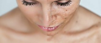
Freckles are a natural feature of the skin and form more often in people with fair skin and hair, green or blue eyes. These formations look like dark brown, gray or red dots and appear most often after exposure to the sun. In most cases, they do not require treatment and do not develop into malignant tumors.
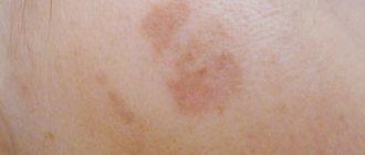
Chloasma looks like spots of light brown or dark color, with a clear, uneven outline. They form most often on the face and hands, after hormonal disruptions, during pregnancy, and menopause.
Melasma can occur on the skin under the influence of ultraviolet radiation, when taking combined oral contraceptives. Senile hyperpigmentation or lentigo is an age-related change in the skin in the form of dark spots of different shapes and sizes.
Melasma is a symptom of endocrine system diseases, such as Graves' disease, pituitary insufficiency, and tumor processes. Berlocc's dermatitis is pigment deposits in the form of a mesh, which occurs as a reaction to cosmetics and some medications.
There is also a distinction between congenital and acquired hyperpigmentation. Congenital pigmentation refers to skin features such as freckles, moles and birthmarks. It can be noted that hyperpigmentation is divided into primary, which occurs on unchanged skin, and secondary, which occurs after allergic or infectious rashes.
Depending on the location of the skin change, it can be isolated - only on one part of the body, or widespread - observed in all areas. Facial pigmentation is also classified into mandibular (in the jaw area), centrofacial (located in the middle of the face), nasolabial (in the area of the nasolabial triangle).
Causes
The spot on the leg may be a mole or freckle. Increased pigmentation of the lower extremities is the result of melanin accumulation. And if it appears under the influence of ultraviolet radiation, then this is perhaps the safest cause of hyperpigmentation of the legs.
Dark spots can also appear as a result of ruptured capillaries. In this case, the area is small and does not require therapeutic measures. In other cases, the causes of pigmentation on the legs or groin lie in one or another pathology. Below is a list of diseases that can cause skin pigmentation on the legs.
- Vascular lesions as a result of diabetes, atherosclerosis and varicose veins.
- Neurofibromatosis. This is the name given to numerous inclusions of a milky-brown hue. They appear for genetic reasons.
- Chronic dermatitis. It is caused by wearing tight clothing, allergies to cosmetic products, fabrics. Stasis dermatitis develops as a result of varicose veins of the deep veins. Areas of darkening on the skin itch, become rough, and become rough.
- Dark areas may appear due to cirrhosis or fibrosis of the liver tissue.
- A dark spot on the skin may be a sign of developing carcinoma. Melanomas on the sole are especially dangerous.
- Some pathologies of the cardiac and vascular system can cause brown spots on the skin of the legs.
- The skin also darkens with vitamin deficiency - a lack of B vitamins.
- If redness appears in the area of the little finger or thumb, then this is the first sign of the development of psoriasis. With this disease, pink or reddish pigmentation appears on the feet and ankles.
Age spots on women's legs
Some women may experience brown spots on their legs while pregnant. The cause of pigmentation on the legs is hormonal changes. After childbirth, skin color returns to normal.
Interesting! Only women may experience characteristic skin changes in the form of chloasma. This is focal hyperpigmentation of the skin. There is no treatment for chloasma: all cosmetic measures are limited to reducing the intensity of skin coloring.
Age spots on the legs of men
Pigmentation on the legs in men can develop as a result of Becker's melanosis . It looks like a mole and develops most often in teenagers. In an area of increased pigmentation, increased hair growth may be observed. The cause of Becker's melanosis has not yet been clearly established. If varicose veins are left untreated, pigmentation of the legs below the knee appears in the form of pink and then red spots. In severe cases, their color changes and they become purple, almost black.
Note! The appearance of black spots indicates that a necrotic process is developing in the skin. Without emergency treatment measures (surgery), gangrene may develop.
Pigmentation of legs with varicose veins
Varicose veins spots appear if the disease has reached an advanced stage. Untimely treatment of the pathology leads to the fact that the veins become clogged with blood clots. Because of this, oxygen metabolism in the tissues is disrupted and they experience oxygen starvation. This is one of the reasons for increased pigmentation of the skin on the legs.
Important! Skin pigmentation on the lower leg and other parts of the limbs does not appear in one day. The skin turns red because blood stagnates in the legs. If varicose veins are left untreated, the skin turns brown. When trophism is disrupted, the skin becomes covered with dark areas.
Predisposing factors
- wearing tight shoes;
- constant microtrauma of the skin of the legs;
- hyperhidrosis;
- use of low-quality household chemicals.
Treatment methods for hyperpigmentation
Treatment of hyperpigmentation depends on its cause, symptoms, and the results of additional studies. For each patient, a treatment plan must be drawn up individually, taking into account age and gender, chronic diseases, and medication use.
When identifying the cause of pigmentation, etiotropic treatment must be carried out. In the case of skin cancer, specific chemotherapy and surgery are carried out; in case of hormonal diseases, replacement therapy is carried out.
For dermatological diseases, such as mycoses, viral skin lesions or streptoderma, antibacterial, antiviral or antifungal therapy is carried out.
The treatment regimen uses additional general treatment, such as vitamin and mineral complexes, medications to strengthen the immune system, folic and nicotinic acids.
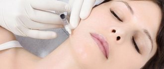
To reduce the appearance of pigmentation, cosmetic procedures may be required, such as chemical peels, ultrasound peels, laser resurfacing, photorejuvenation and ozone therapy.
After performing such hardware treatment methods, it is necessary to give the skin the opportunity to recover. To do this, it is recommended to avoid excessive sun exposure for a week after the procedure, not to use rough scrubs and peels, and to apply sunscreen and moisturizer every day.
To completely eliminate a cosmetic defect, several procedures may be required, with a break of a week to a month. During treatment, it is necessary to avoid direct sunlight on the skin, not to visit solariums, saunas and baths, and protect the skin from mechanical and chemical irritants.
A more gentle means for treating minor pigmentation are chemical peels using fruit and lactic acids, Azalein, and Hydroquinone preparations. These manipulations can be performed more often, the recovery period after them is shorter, but they are less effective than hardware ones.
Between cosmetic procedures, you can use home skin care products, but only with the permission of your doctor.
There are such folk methods of reducing hyperpigmentation - masks using yeast, milk and cream, compresses from potato juice, birch sap, rubbing with celandine juice.
Apply crushed strawberry pulp and a mask of crushed pumpkin seeds. For exfoliation, you can use homemade scrubs and peels made from honey, sugar, and coffee grounds.
It must be remembered that it is undesirable to use hydrogen peroxide or citric acid to whiten skin areas, as they can cause chemical burns, the occurrence of significant skin defects, and the development of an inflammatory process. Filorga cream may be suitable for home use for local skin lightening.
The treatment complex also necessarily includes lifestyle changes, following a gentle hypoallergenic diet, drinking enough liquid, avoiding excessive exposure to the sun, avoiding stress and overexertion.
Types of spots on legs
There are three types of pigmented areas on the legs.
- Leucoderma . It is characterized by decreased pigmentation of the lower extremities, and the skin on the affected areas appears much lighter.
- Melasma . Darkening of the skin is characteristic.
- Blue-gray pigmentation.
These types of disorders are not independent types of pathologies. They indicate that some pathological process is occurring in the body.
Anti-pigmentation cream on the face: which is better, and how to choose the right product
Types of stains:
- mesh;
- marble;
- spotted;
- leprosy;
- lichen;
- keratoses;
- lentigo (age spots);
- post-inflammatory pigmentation;
- warts;
- moles;
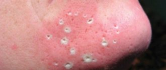
- comedones (or the so-called strawberry legs effect);
- venous stasis (in which the skin becomes dark and even purple over a large area of the leg);
- diabetic spots;
- chronic pigmentary purpura;
- petechiae, or marble hemorrhages;
- Kaposi's sarcoma (caused by herpes virus type eight).
Treatment of age spots on legs
Many patients are interested in how to remove pigmentation on their legs. This can only be done after its cause has been established. Self-medication of discolored legs is dangerous and can cause burns. First of all, you need to contact a dermatologist, phlebologist, or endocrinologist. Women should definitely visit a gynecologist. The causes and treatment of pigmentation on the legs are closely related, and doctors will prescribe a comprehensive examination. After this, it will be possible to select the most effective treatment.
Treatment for hyperpigmentation consists of:
- getting rid of the underlying pathology;
- strengthening the immune system;
- taking antihistamines (most often in the form of an ointment);
- sclerotherapy.
Hyperpigmentation can be removed:
- chemical types of peeling;
- laser;
- whitening creams.
Medicinal treatments
There are medical ways to get rid of hyperpigmentation on the legs. They are selected by the doctor depending on the cause of their occurrence.
- If the cause of the spots is age-related changes, anti-aging drugs with vitamins A and antioxidants are prescribed.
- For fungal pathologies, ointments and creams with a fungicidal effect should be used.
- If a mole has formed, it does not need to be touched. Removal of a nevus occurs only according to a doctor’s indications.
- For age spots on the legs, vitamin complexes with minerals are used.
- If the cause of the spots is varicose veins, take tablets and ointments to strengthen the veins.
- If you have diabetes, you should take antihyperglycemic drugs or insulin (Related: What can you eat if you have diabetes?).
- It is useful to take immunomodulators.
Clinic and diagnosis of pigmentation
The clinical picture of this pathology is varied and depends on the cause of its occurrence. The color of the affected areas of the skin can vary from yellow or light brown to dark and almost black, and the rash can be of any shape.
Pigmented areas are most often located on the face, arms, stomach and back; hyperpigmentation is very rare on the palms, feet and scalp. There are no other complaints or symptoms with normal physiological pigmentation; if pain, itching or burning is observed, you should consult a doctor and conduct additional research.
To make an accurate diagnosis and exclude endocrinological, oncological and other diseases, it is necessary to collect complaints and medical history, and additional research methods. These include a general blood and urine test, biochemical analysis, scatological analysis, ultrasound of the abdominal organs, and chest radiography.
These studies will help discover the cause of pigmentation if it is caused by a certain disease. To exclude oncological pathology, general clinical diagnostic methods, tests for tumor markers, biopsy and histological examination of the affected area are performed. If endocrine disorders are suspected, blood tests are performed for hormones of the pituitary gland, thyroid gland, and adrenal glands. Consultation with an endocrinologist, oncologist, gastroenterologist, and gynecologist for women is mandatory.
Dermatological studies include dermatoscopy, scraping of the affected area of skin, its cytological and histological examination.