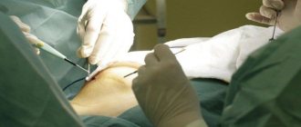
The main options for navel surgery
Have you been trying to heal your JOINTS for many years?
Head of the Institute for the Treatment of Joints: “You will be amazed at how easy it is to cure your joints by taking the product every day for 147 rubles .
Navel surgery is an aesthetic plastic surgery, most often it is performed:
Our readers successfully use Sustalaif to treat joints. Seeing how popular this product is, we decided to bring it to your attention. Read more here...
- Ladies after pregnancy and childbirth. Traditionally, within approximately 6 months after childbirth, the navel returns to normal along with the skin, muscle base of the abdomen and subcutaneous tissue. But, in some cases, the deformation of the navel turns out to be permanent; umbilicoplasty after childbirth can quickly eliminate the defect and restore the woman’s self-confidence.
- Patients after a bad piercing. The reasons for the deformation of the navel in this option will be: incorrect piercing technique, incorrect puncture of the navel ( or navel (Latin umbilicus) - a scar on the anterior abdominal wall remaining after removal of the umbilical cord in a newborn child
), violation of sterility, the body’s tendency to excess formation of connective tissue, allergy to iron objects. - Patients whose navel shape has changed after surgery. Deformation of the navel can be caused by surgical incisions in the area where the navel is located (along the middle strip of the tummy), tension of the skin of the tummy (plastic surgery to tighten the tummy without moving the navel).
- Men and women with acquired features of the umbilical ring structure. This is a bulging navel, formed due to improper handling of the umbilical cord in infancy or helplessness of connective tissues, which leads to the formation of an umbilical hernia and expansion of the umbilical ring.
- Older patients. Over the years, body tissues lose their elasticity, elasticity and sag downwards, age-related ptosis of the skin of the tummy leads to a change in the shape of the navel.
Possible complications
After umbilicoplasty, the patient may experience the following complications:
- Pain and discomfort: nagging pain in the lower abdomen, in the area of the postoperative wound. As a rule, such pain is not intense and can be easily relieved by taking painkillers.
- Hematomas: Postoperative bruises can be extensive, occupying most of the surface of the abdomen. Hematomas usually resolve on their own within 2-3 weeks and do not require additional special treatment.
- Swelling: after navel surgery, significant swelling of the abdominal tissues is possible, due to which the volume of the waist and abdomen may temporarily increase by 1-2 sizes. Most of the swelling goes away after 1-2 weeks, the rest resolves after 1-2 months.
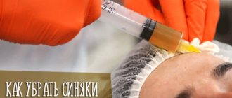
- Infection: occurs due to the doctor’s violation of the rules of asepsis during surgery, which led to infection entering the wound, causing inflammation or purulent melting of tissue. To prevent wound infection, the plastic surgeon prescribes a set of antibiotics, which must be started before surgery and continued after.
- Significant scarring: An overgrowth of connective tissue that results in the formation of rough scars. If in the postoperative period the scars begin to protrude above the surface of the skin and spread wider, then the patient urgently needs to consult a plastic surgeon. Injection of corticosteroid medications can smooth out postoperative scars.
- Formation of an umbilical hernia: a hernia is formed due to a doctor’s violation of the plastic surgery technique, which led to damage to the connective tissue of the umbilical ring. An umbilical hernia can only be removed surgically.
The main and most dangerous complication of an abdominal hernia is its strangulation, in which the patient experiences tissue necrosis and develops anemia. Untimely treatment can lead to peritonitis, which poses a mortal danger to the patient's life.
Main types of hernias
There are three main types ( VIEW: Literally: That which is visible to the eye
) hernias (lat. hernia): umbilical, inguinal and hernia of the snow-white stripe of the tummy.
The most common is a navel hernia .
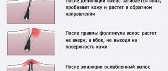
It is also called fumes. In usual cases, when a child is born and there is no longer a need for umbilical cord vessels, the umbilical ring closes and is overgrown with connective tissue.
When the narrowing and closure of the umbilical ring does not occur, then an umbilical hernia develops, which is what we are talking about now.
In most cases, umbilical hernia in children goes away on its own.
An inguinal hernia never goes away on its own; it can only grow. Therefore, she must be operated on immediately. Urgently.
Hernia of the snow-white stripe of the tummy - what is it? We are talking about a ligament in the middle of the tummy. It is mostly small and only looks like a cosmetic flaw. If she doesn’t bother you, you can live with her. The operation consists of 2-3 stitches. In adults it only goes away with local anesthesia. These hernias themselves do not disappear. but they can be operated on when it is comfortable for you.
Why is surgery desirable? Because the bulge of the snow-white stripe of the tummy will spread even more during pregnancy in women and during sports exercises with lifting barbells or weights in boys. These are the individualities of the 3 types of hernial formations - in a nutshell.
Look at the photo of some other types of hernia:
What is an umbilical hernia and for what reasons does it occur in children?
The child, while in the mother's womb, is connected to her by the umbilical cord, through which he receives nutrients for formation and growth. After birth, the umbilical cord is tied up and cut off, and the umbilical cord is no longer needed.
Over time ( the form of physical and mental processes, the condition for the possibility of change
) the umbilical ring (
a round object with a hole inside (example: torus or solid torus)
) is tightened thanks to the muscles of the abdominal cavity. Since the umbilical ring in newborns is weak, from time to time it happens that it does not close completely, and this leads to the bulging of the digestive loop through it.
An umbilical hernia is a condition in which abdominal organs protrude under the skin through the umbilical ring. Most often, the disease is diagnosed in newborns, but it is also observed in one-year-old children and at 6-8 years old.
Mode
But whether the patient is at home or in the hospital, he must be in bed for the first 2-3 days. That is, lie down all the time so that the stitches do not come apart due to an unusual overload for the body.
In some places, on the 3rd day, a person may begin to roll over and stand up. At the same time, it is necessary to realize that it is necessary to exclude any physical overload or fatigue for several days.
The next day after discharge and for a certain period (which is 7-10 days), the patient will have to visit the hospital for dressings. Later, the patient can perform the dressings himself. The nurse assigned to him will teach him this.
A week after the operation ( an action or a combination of actions to achieve a goal
) the patient is prescribed painkillers, medications and physiotherapeutic sessions that will promote rapid primary healing of the operated area.
In older people, after surgery, respiratory failure with tachycardia may occur. This may be a bad sign, which is best reported to the operating doctor.
How to avoid complications of suture healing?
The most common complications during healing are:
- Suppuration.
- Seams coming apart.
Suppuration
If a tumor, hard nodule or lump appears in the area of the sutures, this indicates possible purulent inflammation. It is often accompanied by pain. If leukocytosis is detected (an increase in the number of leukocytes to more than 9 million in 1 ml of blood), it is worth talking with a high degree of confidence about the development of a purulent complication. To eliminate this dangerous complication, you need to examine the wound area daily for swelling and pain.
Postoperative sutures must be properly processed
Seam divergence
The first reason for this is non-compliance with the recommended physical activity regimen. In order for the suture to heal on time, it is necessary to avoid heavy static and dynamic loads for at least a month. The best means of prevention is wearing special bandages. They are sold in regular pharmacies or orthopedic salons.
It is necessary to monitor the food consumed and water intake. Coarse fiber, fatty foods and excess carbohydrates should be excluded during the postoperative recovery period. These products enhance peristalsis and promote the development of flatulence.
A special group of patients are diabetics. If a patient has diabetes mellitus, all reparative and regenerative abilities are reduced. Therefore, any wounds heal worse. This is why diabetics are given special attention during surgical interventions, including laparoscopy. Compensation of glycemia using oral hypoglycemic therapy or insulin therapy in adequate doses is the key to the normal course of the postoperative period and rapid wound healing.
If the sutures come apart, the wound should be considered infected. Therefore, it is treated with antiseptic solutions every day. In this case, systemic antibiotic therapy is carried out in parallel. The stitches are reapplied. If eventration occurs, laparotomy is possible.
When is surgery to remove an umbilical hernia prescribed?
This difficult disease is cured by a surgeon, and you need to contact him as soon as unpleasant feelings arise. Symptoms of an umbilical hernia include the following:
- pain in the abdomen when coughing or exercising;
- presence of nausea;
- extended umbilical ring.
An umbilical hernia can be diagnosed using several methods:
- Get examined by a specialist.
- Take an x-ray of the stomach and duodenum.
- Do an ultrasound.
- Undergo a gastroscopy function.
- To perform such a function as herniography is an x-ray method that involves introducing a special contrast agent into the abdominal cavity, which allows one to study the hernia.
Umbilical hernias can be of two types: congenital and acquired. Congenital can be found immediately after the birth of the baby. In the area of the navel, where the umbilical cord was, there is a spherical bulge with a wide base, running into the umbilical cord. If the baby screams loudly, the hernial bulge increases. How different congenital or acquired hernias can be can be seen in the video that is shown to clients in a medical institution. How to cure an umbilical hernia? Traditionally, surgical treatment of a hernia is not performed until the age of five. They are trying to eliminate it with the help of massage and healing physical exercise. If nothing helps and the navel does not miniaturize, you have to resort to surgical intervention for a hernia ( a disease in which internal organs protrude out of the cavity they normally occupy through a normally existing or pathologically formed hole in the
).
Postoperative hernia on the abdomen
- Localization and classification
- Why do some patients develop hernias after surgery, while others do not?
- Symptoms
- How is diagnosis carried out?
- What complications are possible with an untreated hernia?
- What should people do if they notice postoperative signs of a hernia?
- Prognosis and prevention
- Video on the topic
A postoperative hernia on the abdomen, or more precisely, in the abdominal wall, is a type of traumatic impact. It appears in the area of the postoperative scar and is located under the skin. In relation to all types of hernias, this type is 6–8%.
According to statistics of complications in the postoperative period, hernias account for up to 5% of all manipulations with opening the abdominal cavity, and when analyzing the course of festering wounds, the figure increases to 10%. Prevention measures depend not only on the type of surgical intervention and the doctor’s skills, but also on the patient’s behavior and compliance with recommendations during the rehabilitation period.
How is diagnostics carried out?
What physical therapy exercises can you do?
A physical therapy complex aimed at speedy recovery of the body after surgery may include basic gymnastic exercises that relax and strengthen the muscles of the abdomen, back and hips. All ab exercises are prohibited.
It is necessary to do similar gymnastics daily, dividing the entire complex into several approaches, each of which lasts 15-20 minutes.
Examples of various exercises are below, but it is better to discuss any exercises with your doctor:
And it must be worn constantly for the entire period of time determined by the attending physician, removing the device only during sleep or rest.
Sutures after surgery: features
Removal of suture threads is scheduled approximately one week after surgery. This procedure causes discomfort and some pain at the suture site. After removing the threads, doctors recommend continuing to wear a bandage or a belt that replaces it for several days. Absolute restoration of muscle tone and all connecting damaged tissues occurs two months after surgery. After this, you can completely stop wearing the bandage and return to your normal rhythm and lifestyle. However, rehabilitation depends on the individual characteristics of the body, and wound healing can occur at different times. The healing time also depends on compliance with the doctor’s instructions, diet and other factors. With any load on the patient’s abdominal area, the stitches may come apart or the internal ligaments and muscles may tear. Therefore, it is recommended to wear the bandage constantly until the muscles are completely restored.
Therefore, it is recommended to wear the bandage constantly until the muscles are completely restored.
How is surgery performed to remove an abdominal hernia?
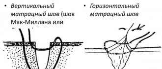
If the contents of the hernial sac are preperitoneal fat, then the operation includes the application of U-shaped sutures. First you need to make sure that inside the hernial orifice there is only abdominal fat and there are no intestinal loops.
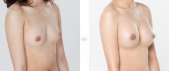
Elimination of diastasis and umbilical hernia (PHOTO AFTER OPERATION)
I finally found a moment to write about my experience in eliminating diastasis and hernia after childbirth. In my case, due to the presence of a hernia, which was rapidly growing, only surgery was indicated; it CANNOT be removed with any exercises. Moreover, it is fraught with strangulation of the hernia, so the first thing I want to say is if the same trouble happens to you after giving birth, SEE A DOCTOR! to a competent surgeon! Unless the muscle diastasis is large, a course of special exercises will help; if there is also a hernia, such exercises are contraindicated in most cases.
So. In order. My son is now one year and 3 months old, the pregnancy was difficult, polyhydramnios due to a large child, natural birth, the belly was huge during pregnancy. HUGE. I’ve always been tall and thin, my abs are almost perfect... I gained a little during pregnancy, but already in the 2nd trimester I realized that my stomach... to put it mildly... in short, I have to work a lot on myself after giving birth. And so it happened - the skin was flabby, covered in small light stretch marks, there was a 2-finger separation of the muscles (lucky, not too big!), and a hernia that was growing... at the time of the operation it was like half a quail egg and began to hurt, pull... when my son turned for a little over a year - I went to the surgeon - the verdict was ONLY OPERATION, it is very dangerous to walk with a hernia, especially since it gave me discomfort and there was a risk of strangulation (despite its small size). It is necessary to install a mesh and sew up the diastasis. I had it done at the clinic named after. Pirogov from St. Petersburg, this pleasure cost 100,000, doctor... the doctor is simply BRAVO. I installed a large enough mesh WITHOUT a vertical incision and a scar. Through the navel. When I looked at photos of similar operations, almost everywhere there was a vertical incision from the navel ((((I was very afraid of this! I don’t want to feel complex later, after all, you can’t disguise a vertical scar and almost always it is visible due to the presence of loose skin, folds form (But I was lucky) On March 31st I had an operation (4 hours, general anesthesia), on April 3rd I was already discharged. About the sensations after the operation... it was just a nightmare on the first day, on the second I was already up on my own. I spent a week at home almost constantly, it was pretty bad . I think the reason is still in the long general anesthesia. To do it beautifully, the doctors had to increase the operation time to 4 hours. After 2 weeks I was already running around the house after the baby))) But it was very difficult - you couldn’t pick him up! Nothing heavier 2 Do not lift kg at all for 2 months and MUST wear a special tight bandage. I still wear it during the day with breaks at night, although in theory it can be removed after 2 months. When pressing on the navel, there is now still a slight discomfort in one place with sudden movements I feel the mesh, BUT ALL THESE INCONVENIENCES ARE WORTH IT. The stitches were removed on the 10th day painlessly, there were only a few of them, only in the navel area. Now there is no fear of strangulating a hernia, and you can see the cosmetic effect in the photo! My waist has lost more than 4 centimeters, despite the fact that I have gained 3 kg after the operation)))) The navel is not quite perfect yet - there are small folds, but this is scar tissue - it should smooth out. At least I hope that my tummy will be completely smooth soon! I wish good luck to everyone who has encountered this problem. Decide to have surgery, if it is really necessary, let everything go well))
When is the stitch removed?
The time for removing suture material is individual for each specific case. It all depends on the severity of the condition, the regenerative ability of the skin and a host of other factors.
The average time for uncomplicated sutures is up to 2 weeks. If healing is good without discomfort or signs of inflammation, they can be removed after 8–10 days.
To prevent suppuration, brilliant green or other antiseptics are used
After the stitches are removed, the wound area is treated at home. To do this, use iodine-containing or any other antiseptics that the doctor recommends. After surgery, the scars are initially purplish-red. Then, over time, they become paler and completely light.
To make the marks less noticeable after laparoscopy, you can use physiotherapeutic methods of influence. This is UHF, electrophoresis with potassium iodide or lidase. But this should be done only after consultation with the doctor.
Umbilical hernia in adults - reviews of the operation, symptoms and treatment
Methods of performing surgery to remove an umbilical hernia: laparoscopic, open, hernioplasty, laser removal. ⭐️⭐️ ⭐️ ⭐️ ⭐️ Duration of surgery, rehabilitation and possible complications. Forecasts and patient reviews
Read advice from our experts
Symptoms of an umbilical hernia are:
- compaction or protrusion in the navel area;
- expansion of the umbilical ring;
- pain in the middle of the abdomen during physical activity, coughing;
- nausea.
The disease can occur at any age in both men and women. But more often, umbilical hernia occurs in infants and people over 40 years of age.
Weakness of connective tissue and abdominal muscles predisposes to the appearance of a hernia in adults. Protrusion can occur with increased physical activity, heavy lifting, or chronic diseases accompanied by coughing. Excess weight, constipation, pregnancy, and ascites (accumulation of fluid in the abdominal cavity) also contribute to the formation of a hernia.
An umbilical hernia in infants can be congenital (due to improper formation of organs in the womb) or acquired. The latter occurs with frequent increases in pressure inside the peritoneum due to crying and slow healing of the umbilical ring. In some cases, infants' hernias are repaired non-surgically - with the help of massage.
The primary examination of a hernia consists of a visual examination of the patient by a general practitioner. Inspection and palpation are carried out in a horizontal and vertical position. If a hernia is detected, the doctor prescribes further examinations:
- Ultrasound of the abdominal organs;
- herniography – diagnosis using a contrast agent injected into the peritoneum;
- FGDS (for the study of a complex of diseases of the gastrointestinal tract);
- computed tomography.
To clarify the shape and size of the hernia, ultrasound diagnostics is sufficient. However, for a more detailed study and identification of concomitant diseases, additional methods are used.
There are a number of contraindications to surgery for umbilical hernia in adults and children. Therefore, it is necessary to carry out examinations in advance, the results of which may force the doctor to reschedule or cancel the intervention. Preparation before surgery to remove an umbilical hernia includes the following tests:
- ECG;
- general blood analysis;
- Analysis of urine;
- coagulogram;
- fluorography;
- tests for HIV, hepatitis, syphilis.
Contraindications to surgery to remove an umbilical hernia are:
- severe cardiovascular disease, recent heart attack or stroke;
- pregnancy, especially 2-3 trimester;
- old age – from 80 years and older;
- cirrhosis of the liver;
- renal failure;
- diabetes;
- varicose veins of the esophagus;
- infectious diseases in the acute stage;
- blood clotting disorder.
Hernioplasty is considered a simple operation, especially for small hernias. But even in emergency cases, when the hernial sac is strangulated or grows, surgical intervention, as a rule, goes well and is successful for the patient.
Removal of an umbilical hernia in adults and children can be performed using the traditional method (using tissue tension, without the use of an implant) or with the installation of an endoprosthesis . The implant is a mesh that strengthens the weakened part of the peritoneum and promotes its overgrowth with new tissue. Endoprostheses are made from different materials. Some of them dissolve over time, leaving behind their own tissues that have grown on top. Others remain in place and can be removed if necessary. Surgery using an endoprosthesis helps reduce the risk of relapse by strengthening the abdominal wall.
Laparoscopic method
About 25 years ago, it became possible to use a gentle method for removing an umbilical hernia - laparoscopic. Now this is the main and safest way to treat hernias. The operation is performed through small incisions, into one of which a camera is placed, and into the other - surgical instruments. Due to its low invasiveness, the risk of adhesions is reduced and the postoperative period is shortened. Sutures after laparoscopy are practically invisible. Laparoscopic surgery for umbilical hernia in women is performed during the intermenstrual period. With laparoscopy, both the tension method of closing the hernial orifice and the installation of a mesh implant are possible.
Single-port laparoscopy or the single-incision SILS method has been used relatively recently and has quickly gained the sympathy of both surgeons and patients. The disadvantage of this method is that the puncture hole is much larger than the standard ones.
Hernioplasty using Sapezhko, Lexer or Mayo methods
These are traditional methods of hernioplasty, which involve closing the hernial orifice using tissue tension. There are some differences in the course of operations and indications for them.
The Mayo method is more often used in overweight people. Two crescent-shaped incisions are made around the navel to capture excess fat deposits. Then the hernial sac is cut, adhesions and necrotic tissue are removed, and the internal organs are set into the peritoneum. The edges of the hernial sac are sutured horizontally. The aponeurosis (the so-called plate of connective tissue that gives strength to the abdominal wall) is sutured.
During surgery using the Sapezhko method, incisions are made in the form of vertical lines. After placing the organs in the abdominal cavity, layer-by-layer stitching of tissue is performed. Otherwise, the procedure is almost the same as the Mayo method.
The Lexer method is suitable for cases where the navel and hernial sac are inseparable. If necessary (large size of the hernia), the navel is removed during the operation. The doctor makes a crescent-shaped incision around the tumor. When the hernial sac fuses with the navel, it can be difficult to release its contents. The bottom or neck of the hernial sac is opened, and the organs are placed in the abdominal cavity. A suture is placed on the neck, and the sac itself is cut off. The hernial orifice is closed using the Lexer method by applying a silk suture to the aponeurosis around the ring and several sutures to the rectus abdominis muscles. After this, the skin flap cut out at the beginning of the operation is returned to its place and secured with interrupted sutures.
Postoperative period and rehabilitation for umbilical hernia
The main cause of this disease is the weakening of the umbilical ring and the formation of a hole in the anterior abdominal wall, through which the internal organs, the intestines and the greater omentum, emerge under the skin. An umbilical hernia develops more often in women, which is associated with stretching of the umbilical ring during pregnancy. The very appearance of a hernia indicates that the organs have changed their location, and their normal mutual pressure on each other has been disrupted. This leads to disruption of the functions of all organs involved in the formation of a hernia. An umbilical hernia must be treated. Without effective treatment, even a small hernia significantly impedes the normal functioning of organs. An umbilical hernia is often accompanied by chronic constipation.
Removal of inguinal hernia Removal of umbilical hernia Cost of removal of umbilical hernia in adults Treatment of umbilical hernia in adults Treatment of umbilical hernia without surgery Bandage for umbilical hernia Umbilical hernia in women Diastasis of the anterior abdominal wall Postoperative ventral hernia Spigelian line hernia Alba linea hernia Femoral hernia Surgery to remove the umbilical hernias in Moscow Treatment and surgery to remove an inguinal hernia using laparoscopy Stretch marks on the abdomen after childbirth How long does it take for the belly to go away after childbirth Plank for diastasis More articles.